

domains as is the case of PICK1 and MUPP1, respectively. In some
cases, two PDZ domains can be located in close proximity to each
other inside the same protein, such that they are arranged into a
PDZ tandem that may act as a single functional unit [4, 6].
PDZ domains are normally found together with other protein–
protein interaction domains in the same protein. For example,
proteins from the membrane-associated guanylate kinase
(MAGUK) include an SH3 module, multiple PDZ domains, and
a guanylate kinase domain which is inactive. The presence of different protein–protein interacting domains allows proteins containing
PDZ domains to bind many proteins at the same time and act as a
scaffold. This is why they participate in a wide variety of biological
processes such as cell polarity regulation, cell–cell interactions, cell
migration, proliferation and survival, intracellular transport, signal
transduction, or protein arrangement [5, 7].
PDZ domains mainly interact with a short stretch of amino acid
residues arranged in a specific manner called PDZ-binding-motifs
(PBMs). However, they may also interact with other PDZ domains
or even phospholipids [8, 9]. PBMs are specific sequences usually
located in the last amino acids of the protein. Although controversial, PDZ domains can be classified in three different groups,
according to the PBM sequence they interact with: Class I PDZ
domains recognize the motif X-S/T-X-Φ-COOH (where X could
be any amino acid and Φ is a hydrophobic amino acid (normally V,
I, or L), class II PDZs bind the sequence X-Φ-S-Φ-COOH, and
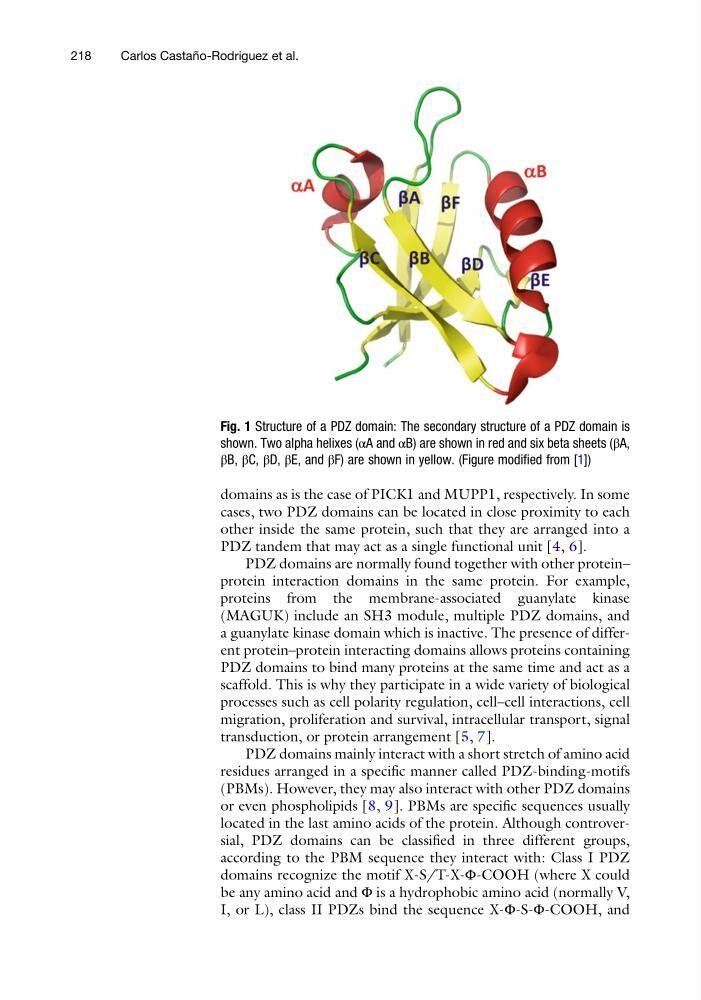
Fig. 1 Structure of a PDZ domain: The secondary structure of a PDZ domain is
shown. Two alpha helixes (αA and αB) are shown in red and six beta sheets (βA,
βB, βC, βD, βE, and βF) are shown in yellow. (Figure modified from [1])
218
Carlos Castan ˜ o-Rodriguez et al.
cases, two PDZ domains can be located in close proximity to each
other inside the same protein, such that they are arranged into a
PDZ tandem that may act as a single functional unit [4, 6].
PDZ domains are normally found together with other protein–
protein interaction domains in the same protein. For example,
proteins from the membrane-associated guanylate kinase
(MAGUK) include an SH3 module, multiple PDZ domains, and
a guanylate kinase domain which is inactive. The presence of different protein–protein interacting domains allows proteins containing
PDZ domains to bind many proteins at the same time and act as a
scaffold. This is why they participate in a wide variety of biological
processes such as cell polarity regulation, cell–cell interactions, cell
migration, proliferation and survival, intracellular transport, signal
transduction, or protein arrangement [5, 7].
PDZ domains mainly interact with a short stretch of amino acid
residues arranged in a specific manner called PDZ-binding-motifs
(PBMs). However, they may also interact with other PDZ domains
or even phospholipids [8, 9]. PBMs are specific sequences usually
located in the last amino acids of the protein. Although controversial, PDZ domains can be classified in three different groups,
according to the PBM sequence they interact with: Class I PDZ
domains recognize the motif X-S/T-X-Φ-COOH (where X could
be any amino acid and Φ is a hydrophobic amino acid (normally V,
I, or L), class II PDZs bind the sequence X-Φ-S-Φ-COOH, and
Fig. 1 Structure of a PDZ domain: The secondary structure of a PDZ domain is
shown. Two alpha helixes (αA and αB) are shown in red and six beta sheets (βA,
βB, βC, βD, βE, and βF) are shown in yellow. (Figure modified from [1])
218
Carlos Castan ˜ o-Rodriguez et al.