

characteristic secondary structures such as α-helices, β-sheets,
turns, and disordered sections with specific Φ, Ψ dihedral angles
and H-bond patterns affecting the CD spectrum. Numerous algorithms have been developed for the quantitative estimation of the
secondary structure composition from the CD spectra with a good
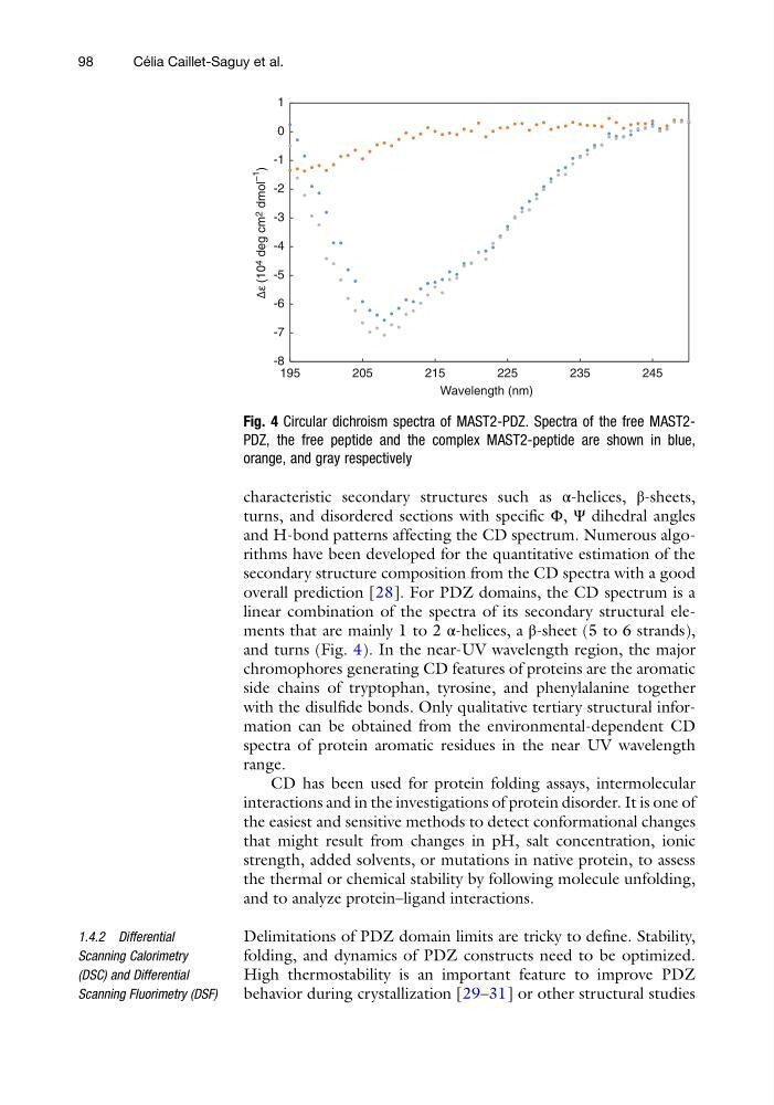
overall prediction [28]. For PDZ domains, the CD spectrum is a
linear combination of the spectra of its secondary structural elements that are mainly 1 to 2 α-helices, a β-sheet (5 to 6 strands),
and turns (Fig. 4). In the near-UV wavelength region, the major
chromophores generating CD features of proteins are the aromatic
side chains of tryptophan, tyrosine, and phenylalanine together
with the disulfide bonds. Only qualitative tertiary structural information can be obtained from the environmental-dependent CD
spectra of protein aromatic residues in the near UV wavelength
range.
CD has been used for protein folding assays, intermolecular
interactions and in the investigations of protein disorder. It is one of
the easiest and sensitive methods to detect conformational changes
that might result from changes in pH, salt concentration, ionic
strength, added solvents, or mutations in native protein, to assess
the thermal or chemical stability by following molecule unfolding,
and to analyze protein–ligand interactions.
1.4.2 Differential
Scanning Calorimetry
(DSC) and Differential
Scanning Fluorimetry (DSF)
Delimitations of PDZ domain limits are tricky to define. Stability,
folding, and dynamics of PDZ constructs need to be optimized.
High thermostability is an important feature to improve PDZ
behavior during crystallization [29–31] or other structural studies
-8
-7
-6
-5
-4
-3
-2
-1
0
1
195
205
215
225
235
245
Wavelength (nm)
Δε (10 4
deg cm 2
dmol
–1
)
Fig. 4 Circular dichroism spectra of MAST2-PDZ. Spectra of the free MAST2PDZ, the free peptide and the complex MAST2-peptide are shown in blue,
orange, and gray respectively
98
Ce ´ lia Caillet-Saguy et al.
turns, and disordered sections with specific Φ, Ψ dihedral angles
and H-bond patterns affecting the CD spectrum. Numerous algorithms have been developed for the quantitative estimation of the
secondary structure composition from the CD spectra with a good
overall prediction [28]. For PDZ domains, the CD spectrum is a
linear combination of the spectra of its secondary structural elements that are mainly 1 to 2 α-helices, a β-sheet (5 to 6 strands),
and turns (Fig. 4). In the near-UV wavelength region, the major
chromophores generating CD features of proteins are the aromatic
side chains of tryptophan, tyrosine, and phenylalanine together
with the disulfide bonds. Only qualitative tertiary structural information can be obtained from the environmental-dependent CD
spectra of protein aromatic residues in the near UV wavelength
range.
CD has been used for protein folding assays, intermolecular
interactions and in the investigations of protein disorder. It is one of
the easiest and sensitive methods to detect conformational changes
that might result from changes in pH, salt concentration, ionic
strength, added solvents, or mutations in native protein, to assess
the thermal or chemical stability by following molecule unfolding,
and to analyze protein–ligand interactions.
1.4.2 Differential
Scanning Calorimetry
(DSC) and Differential
Scanning Fluorimetry (DSF)
Delimitations of PDZ domain limits are tricky to define. Stability,
folding, and dynamics of PDZ constructs need to be optimized.
High thermostability is an important feature to improve PDZ
behavior during crystallization [29–31] or other structural studies
-8
-7
-6
-5
-4
-3
-2
-1
0
1
195
205
215
225
235
245
Wavelength (nm)
Δε (10 4
deg cm 2
dmol
–1
)
Fig. 4 Circular dichroism spectra of MAST2-PDZ. Spectra of the free MAST2PDZ, the free peptide and the complex MAST2-peptide are shown in blue,
orange, and gray respectively
98
Ce ´ lia Caillet-Saguy et al.