

Processes 2018, 6, 134
on the LDLR gene for SREBP to bind to and x h is the number of binding sites on the HMGCR gene to
which SREBP binds.
The mechanisms of LDL and VLDL endocytosis and the subsequent processing of cholesterol and
receptors, is given by the following
W
d ¯
l E
d ¯
t
= −¯ α L ¯
r f ¯
l E + ¯
α −L ¯
l RB + W( ¯
χ v ¯
v E ),
(11)
d ¯
l RB
d ¯
t
= ¯
α L ¯
r f ¯
l E − ¯
α −L ¯
l RB − ¯
β L ¯
l RB ,
(12)
d ¯
l I
d ¯
t
= ¯
β L ¯
l RB − ¯
γ L ¯
l I ,
(13)
W
d ¯
v E
d ¯
t
= −¯ α v ¯
r f ¯
v E + ¯
α −v ¯
v RB + W(− ¯
χ v ¯
v E + ¯
ω),
(14)
d ¯
v RB
d ¯
t
= ¯
α v ¯
r f ¯
v E − ¯
α −v ¯
v RB − ¯
β v ¯
v RB ,
(15)
d ¯
v I
d ¯
t
= ¯
β v ¯
v RB − ¯
γ v ¯
v I ,
(16)
d¯ r f
d ¯
t
= ¯
γ r ¯
r I − P ¯
β 0 ¯
r
P
f − ˜
m l ¯
β L ¯
l RB − m l ¯
α L ¯
l E ¯
r f + m l ¯
α −L ¯
l RB − ˜
m v ¯
β v ¯
v RB − m v ¯
α v ¯
v E ¯
r f
+m v ¯
α −v ¯
v RB ,
(17)
d¯ r I
d ¯
t
= ¯
μ r ¯
m r − ¯
γ r ¯
r I + Pf ¯
β 0 ¯
r
P
f + f (m l + ˜
m l ) ¯
β L ¯
l RB + f (m v + ˜
m v ) ¯
β v ¯
v RB ,
(18)
d ¯
c
d ¯
t
= R
chol
L
¯
γ L ¯
l I + R
chol
v
¯
γ v ¯
v I + ¯
μ c ¯
h − ¯
δ c ¯
c + J(x c ¯
κ −c ¯
c b − x c ¯
κ c ¯
c
xc ¯
s),
(19)
d ¯
c b
d ¯
t
=( ¯
κ c ¯
c
xc ¯
s − ¯
κ −c ¯
c b ),
(20)
where the concentration of free LDL surrounding the cell is ¯
l E , ¯
l RB is receptor bound LDL, ¯
l I is
internalised LDL, ¯
v E is free VLDL surrounding the cell, ¯
v RB is receptor bound VLDL, ¯
v I is internalised
VLDL, ¯
r f are free unbound receptors, ¯
r I are internalised receptors, ¯
c is cholesterol and ¯
c b is the SREBP-2
cholesterol bound complex.
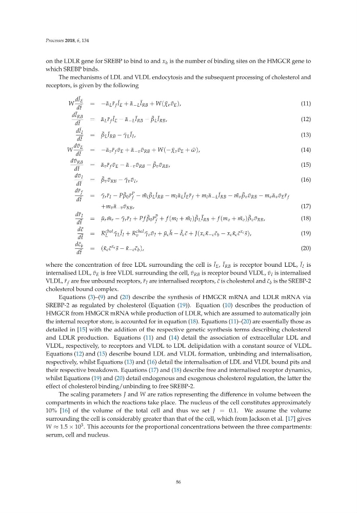
Equations (3)–(9) and (20) describe the synthesis of HMGCR mRNA and LDLR mRNA via
SREBP-2 as regulated by cholesterol (Equation (19)). Equation (10) describes the production of
HMGCR from HMGCR mRNA while production of LDLR, which are assumed to automatically join
the internal receptor store, is accounted for in equation (18). Equations (11)–(20) are essentially those as
detailed in [15] with the addition of the respective genetic synthesis terms describing cholesterol
and LDLR production. Equations (11) and (14) detail the association of extracellular LDL and
VLDL, respectively, to receptors and VLDL to LDL delipidation with a constant source of VLDL.
Equations (12) and (15) describe bound LDL and VLDL formation, unbinding and internalisation,
respectively, whilst Equations (13) and (16) detail the internalisation of LDL and VLDL bound pits and
their respective breakdown. Equations (17) and (18) describe free and internalised receptor dynamics,
whilst Equations (19) and (20) detail endogenous and exogenous cholesterol regulation, the latter the
effect of cholesterol binding/unbinding to free SREBP-2.
The scaling parameters J and W are ratios representing the difference in volume between the
compartments in which the reactions take place. The nucleus of the cell constitutes approximately
10% [16] of the volume of the total cell and thus we set J = 0.1. We assume the volume
surrounding the cell is considerably greater than that of the cell, which from Jackson et al. [17] gives
W ≈ 1.5 × 10 3 . This accounts for the proportional concentrations between the three compartments:
serum, cell and nucleus.
86
on the LDLR gene for SREBP to bind to and x h is the number of binding sites on the HMGCR gene to
which SREBP binds.
The mechanisms of LDL and VLDL endocytosis and the subsequent processing of cholesterol and
receptors, is given by the following
W
d ¯
l E
d ¯
t
= −¯ α L ¯
r f ¯
l E + ¯
α −L ¯
l RB + W( ¯
χ v ¯
v E ),
(11)
d ¯
l RB
d ¯
t
= ¯
α L ¯
r f ¯
l E − ¯
α −L ¯
l RB − ¯
β L ¯
l RB ,
(12)
d ¯
l I
d ¯
t
= ¯
β L ¯
l RB − ¯
γ L ¯
l I ,
(13)
W
d ¯
v E
d ¯
t
= −¯ α v ¯
r f ¯
v E + ¯
α −v ¯
v RB + W(− ¯
χ v ¯
v E + ¯
ω),
(14)
d ¯
v RB
d ¯
t
= ¯
α v ¯
r f ¯
v E − ¯
α −v ¯
v RB − ¯
β v ¯
v RB ,
(15)
d ¯
v I
d ¯
t
= ¯
β v ¯
v RB − ¯
γ v ¯
v I ,
(16)
d¯ r f
d ¯
t
= ¯
γ r ¯
r I − P ¯
β 0 ¯
r
P
f − ˜
m l ¯
β L ¯
l RB − m l ¯
α L ¯
l E ¯
r f + m l ¯
α −L ¯
l RB − ˜
m v ¯
β v ¯
v RB − m v ¯
α v ¯
v E ¯
r f
+m v ¯
α −v ¯
v RB ,
(17)
d¯ r I
d ¯
t
= ¯
μ r ¯
m r − ¯
γ r ¯
r I + Pf ¯
β 0 ¯
r
P
f + f (m l + ˜
m l ) ¯
β L ¯
l RB + f (m v + ˜
m v ) ¯
β v ¯
v RB ,
(18)
d ¯
c
d ¯
t
= R
chol
L
¯
γ L ¯
l I + R
chol
v
¯
γ v ¯
v I + ¯
μ c ¯
h − ¯
δ c ¯
c + J(x c ¯
κ −c ¯
c b − x c ¯
κ c ¯
c
xc ¯
s),
(19)
d ¯
c b
d ¯
t
=( ¯
κ c ¯
c
xc ¯
s − ¯
κ −c ¯
c b ),
(20)
where the concentration of free LDL surrounding the cell is ¯
l E , ¯
l RB is receptor bound LDL, ¯
l I is
internalised LDL, ¯
v E is free VLDL surrounding the cell, ¯
v RB is receptor bound VLDL, ¯
v I is internalised
VLDL, ¯
r f are free unbound receptors, ¯
r I are internalised receptors, ¯
c is cholesterol and ¯
c b is the SREBP-2
cholesterol bound complex.
Equations (3)–(9) and (20) describe the synthesis of HMGCR mRNA and LDLR mRNA via
SREBP-2 as regulated by cholesterol (Equation (19)). Equation (10) describes the production of
HMGCR from HMGCR mRNA while production of LDLR, which are assumed to automatically join
the internal receptor store, is accounted for in equation (18). Equations (11)–(20) are essentially those as
detailed in [15] with the addition of the respective genetic synthesis terms describing cholesterol
and LDLR production. Equations (11) and (14) detail the association of extracellular LDL and
VLDL, respectively, to receptors and VLDL to LDL delipidation with a constant source of VLDL.
Equations (12) and (15) describe bound LDL and VLDL formation, unbinding and internalisation,
respectively, whilst Equations (13) and (16) detail the internalisation of LDL and VLDL bound pits and
their respective breakdown. Equations (17) and (18) describe free and internalised receptor dynamics,
whilst Equations (19) and (20) detail endogenous and exogenous cholesterol regulation, the latter the
effect of cholesterol binding/unbinding to free SREBP-2.
The scaling parameters J and W are ratios representing the difference in volume between the
compartments in which the reactions take place. The nucleus of the cell constitutes approximately
10% [16] of the volume of the total cell and thus we set J = 0.1. We assume the volume
surrounding the cell is considerably greater than that of the cell, which from Jackson et al. [17] gives
W ≈ 1.5 × 10 3 . This accounts for the proportional concentrations between the three compartments:
serum, cell and nucleus.
86