

Processes 2018, 6,38
•
DNA standards: ~300 µg/mL stock solution calf thymus DNA (Sigma D1501, Merck KGaA,
St. Louis, MO, USA), stored at 4 ◦ C. (Concentration was measured with a NanoDrop
1000 spectrophotometer (Wilmington, DE, USA) and was verified after several days and again
after several weeks to ensure a stable concentration.) Prepare a 100 µg/mL working stock solution
with standard buffer. Dilute the working stock into a standard series with standard buffer.
•
Hoechst reagent: Prepare a 200 µg/mL intermediate Hoechst stock from 10 mg/mL stock solution
(Biotium 40044, Fremont, CA, USA). Prepare a 1 µg/mL Hoechst working stock fresh daily from
the intermediate stock with assay buffer. Store solutions at 4 ◦ C wrapped in aluminum foil to
protect from light.
Note: all solutions were prepared using nuclease-free water.
5.2.2. Assay
(1) Re-suspend cell pellet to 50 µL total volume in nuclease-free water in a 2-mL Eppendorf tube.
(2) Add 50 µL of alkali extraction solution.
(3) Incubate at 37 ◦ C for 3 h (VWR analog heat block).
(4) Dilute to 2 mL total volume by adding 1.9 mL assay buffer.
(5) Transfer to a 15-mL polypropylene centrifuge tube and centrifuge (3400 rpm, 30 min, 4 ◦ C).
(6) Aliquot 295 µL of Hoechst working reagent in a clear-bottom black 96-well plate (Corning 3603,
Corning, NY, USA).
(7) Add 50 µL of sample to the well (manual mixing via pipette is unnecessary as the plate reader
mixes by shaking).
(8) Use a fluorescent plate reader (Synergy HT, Gen5 software, BioTek, Winooski, VT, USA) to read
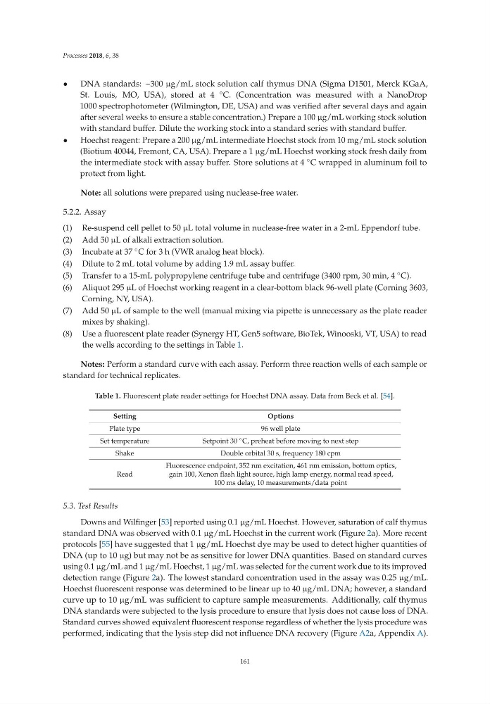
the wells according to the settings in Table 1.
Notes: Perform a standard curve with each assay. Perform three reaction wells of each sample or
standard for technical replicates.
Table 1. Fluorescent plate reader settings for Hoechst DNA assay. Data from Beck et al. [54].
Setting
Options
Plate type
96 well plate
Set temperature
Setpoint 30 ◦ C, preheat before moving to next step
Shake
Double orbital 30 s, frequency 180 cpm
Read
Fluorescence endpoint, 352 nm excitation, 461 nm emission, bottom optics,
gain 100, Xenon flash light source, high lamp energy, normal read speed,
100 ms delay, 10 measurements/data point
5.3. Test Results
Downs and Wilfinger [53] reported using 0.1 µg/mL Hoechst. However, saturation of calf thymus
standard DNA was observed with 0.1 µg/mL Hoechst in the current work (Figure 2a). More recent
protocols [55] have suggested that 1 µg/mL Hoechst dye may be used to detect higher quantities of
DNA (up to 10 µg) but may not be as sensitive for lower DNA quantities. Based on standard curves
using 0.1 µg/mL and 1 µg/mL Hoechst, 1 µg/mL was selected for the current work due to its improved
detection range (Figure 2a). The lowest standard concentration used in the assay was 0.25 µg/mL.
Hoechst fluorescent response was determined to be linear up to 40 µg/mL DNA; however, a standard
curve up to 10 µg/mL was sufficient to capture sample measurements. Additionally, calf thymus
DNA standards were subjected to the lysis procedure to ensure that lysis does not cause loss of DNA.
Standard curves showed equivalent fluorescent response regardless of whether the lysis procedure was
performed, indicating that the lysis step did not influence DNA recovery (Figure A2a, Appendix A).
161
•
DNA standards: ~300 µg/mL stock solution calf thymus DNA (Sigma D1501, Merck KGaA,
St. Louis, MO, USA), stored at 4 ◦ C. (Concentration was measured with a NanoDrop
1000 spectrophotometer (Wilmington, DE, USA) and was verified after several days and again
after several weeks to ensure a stable concentration.) Prepare a 100 µg/mL working stock solution
with standard buffer. Dilute the working stock into a standard series with standard buffer.
•
Hoechst reagent: Prepare a 200 µg/mL intermediate Hoechst stock from 10 mg/mL stock solution
(Biotium 40044, Fremont, CA, USA). Prepare a 1 µg/mL Hoechst working stock fresh daily from
the intermediate stock with assay buffer. Store solutions at 4 ◦ C wrapped in aluminum foil to
protect from light.
Note: all solutions were prepared using nuclease-free water.
5.2.2. Assay
(1) Re-suspend cell pellet to 50 µL total volume in nuclease-free water in a 2-mL Eppendorf tube.
(2) Add 50 µL of alkali extraction solution.
(3) Incubate at 37 ◦ C for 3 h (VWR analog heat block).
(4) Dilute to 2 mL total volume by adding 1.9 mL assay buffer.
(5) Transfer to a 15-mL polypropylene centrifuge tube and centrifuge (3400 rpm, 30 min, 4 ◦ C).
(6) Aliquot 295 µL of Hoechst working reagent in a clear-bottom black 96-well plate (Corning 3603,
Corning, NY, USA).
(7) Add 50 µL of sample to the well (manual mixing via pipette is unnecessary as the plate reader
mixes by shaking).
(8) Use a fluorescent plate reader (Synergy HT, Gen5 software, BioTek, Winooski, VT, USA) to read
the wells according to the settings in Table 1.
Notes: Perform a standard curve with each assay. Perform three reaction wells of each sample or
standard for technical replicates.
Table 1. Fluorescent plate reader settings for Hoechst DNA assay. Data from Beck et al. [54].
Setting
Options
Plate type
96 well plate
Set temperature
Setpoint 30 ◦ C, preheat before moving to next step
Shake
Double orbital 30 s, frequency 180 cpm
Read
Fluorescence endpoint, 352 nm excitation, 461 nm emission, bottom optics,
gain 100, Xenon flash light source, high lamp energy, normal read speed,
100 ms delay, 10 measurements/data point
5.3. Test Results
Downs and Wilfinger [53] reported using 0.1 µg/mL Hoechst. However, saturation of calf thymus
standard DNA was observed with 0.1 µg/mL Hoechst in the current work (Figure 2a). More recent
protocols [55] have suggested that 1 µg/mL Hoechst dye may be used to detect higher quantities of
DNA (up to 10 µg) but may not be as sensitive for lower DNA quantities. Based on standard curves
using 0.1 µg/mL and 1 µg/mL Hoechst, 1 µg/mL was selected for the current work due to its improved
detection range (Figure 2a). The lowest standard concentration used in the assay was 0.25 µg/mL.
Hoechst fluorescent response was determined to be linear up to 40 µg/mL DNA; however, a standard
curve up to 10 µg/mL was sufficient to capture sample measurements. Additionally, calf thymus
DNA standards were subjected to the lysis procedure to ensure that lysis does not cause loss of DNA.
Standard curves showed equivalent fluorescent response regardless of whether the lysis procedure was
performed, indicating that the lysis step did not influence DNA recovery (Figure A2a, Appendix A).
161