

Processes 2018, 6,42
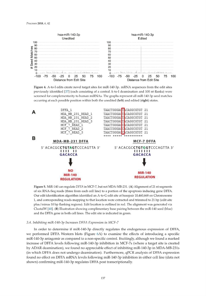
Figure 4. A-to-I edits create novel target sites for miR-140-3p. mRNA sequences from the edit sites
previously identified [37] (each consisting of a central A-to-I deamination and 100 nt flanks) were
screened for complementarity to human miRNAs. The graphs represent all miR-140-3p seed matches
occurring at each possible position within both the unedited (left) and edited (right) states.
Figure 5. MiR-140 can regulate DFFA in MCF-7, but not MDA-MB-231. (A) Alignment of 21 nt segments
of six RNA-Seq reads (three from each cell line) to a portion of the apoptosis inducing gene DFFA.
Our edit identification algorithm identified an A-to-G edit site at basepair 10,460,668 on Chromosome
1, and corresponding reads mapping to that location were extracted and trimmed to 21 bp (edit site
plus/minus 10 bp flanking regions). Edit location is outlined in red. The alignment was generated via
ClustalW [40]. (B) Illustration showing complimentary base pairing between the miR-140 seed (blue)
and the DFFA gene in both cell lines. The edit site is indicated in green.
3.4. Inhibiting miR-140-3p Increases DFFA Expression in MCF-7
In order to determine if miR-140-3p directly regulates the endogenous expression of DFFA,
we performed DFFA Western blots (Figure 6A) to examine the effects of introducing a specific
miR-140-3p antagomir as compared to a non-specific control. Excitingly, although we found a marked
increase of DFFA levels following miR-140-3p inhibition in MCF-7s (where a target site is created
by ADAR deamination), we found no appreciable effect of inhibiting miR-140-3p in MDA-MB-231s
(in which DFFA does not undergo deamination). Furthermore, qPCR analysis of DFFA expression
found no effect on DFFA mRNA levels following miR-140-3p inhibition in either cell line (data not
shown) confirming miR-140-3p regulates DFFA post transcriptionally.
137
Figure 4. A-to-I edits create novel target sites for miR-140-3p. mRNA sequences from the edit sites
previously identified [37] (each consisting of a central A-to-I deamination and 100 nt flanks) were
screened for complementarity to human miRNAs. The graphs represent all miR-140-3p seed matches
occurring at each possible position within both the unedited (left) and edited (right) states.
Figure 5. MiR-140 can regulate DFFA in MCF-7, but not MDA-MB-231. (A) Alignment of 21 nt segments
of six RNA-Seq reads (three from each cell line) to a portion of the apoptosis inducing gene DFFA.
Our edit identification algorithm identified an A-to-G edit site at basepair 10,460,668 on Chromosome
1, and corresponding reads mapping to that location were extracted and trimmed to 21 bp (edit site
plus/minus 10 bp flanking regions). Edit location is outlined in red. The alignment was generated via
ClustalW [40]. (B) Illustration showing complimentary base pairing between the miR-140 seed (blue)
and the DFFA gene in both cell lines. The edit site is indicated in green.
3.4. Inhibiting miR-140-3p Increases DFFA Expression in MCF-7
In order to determine if miR-140-3p directly regulates the endogenous expression of DFFA,
we performed DFFA Western blots (Figure 6A) to examine the effects of introducing a specific
miR-140-3p antagomir as compared to a non-specific control. Excitingly, although we found a marked
increase of DFFA levels following miR-140-3p inhibition in MCF-7s (where a target site is created
by ADAR deamination), we found no appreciable effect of inhibiting miR-140-3p in MDA-MB-231s
(in which DFFA does not undergo deamination). Furthermore, qPCR analysis of DFFA expression
found no effect on DFFA mRNA levels following miR-140-3p inhibition in either cell line (data not
shown) confirming miR-140-3p regulates DFFA post transcriptionally.
137