

Processes 2018, 6, 134
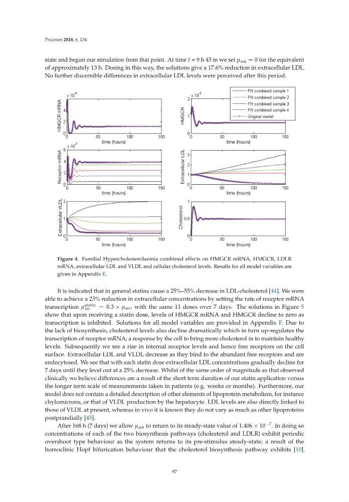
state and begun our simulation from that point. At time t =9h45mwesetμ mh = 0 for the equivalent
of approximately 13 h. Dosing in this way, the solutions give a 17.6% reduction in extracellular LDL.
No further discernible differences in extracellular LDL levels were perceived after this period.
Figure 4. Familial Hypercholesterolaemia combined effects on HMGCR mRNA, HMGCR, LDLR
mRNA, extracellular LDL and VLDL and cellular cholesterol levels. Results for all model variables are
given in Appendix E.
It is indicated that in general statins cause a 25%–55% decrease in LDL-cholesterol [44]. We were
able to achieve a 25% reduction in extracellular concentrations by setting the rate of receptor mRNA
transcription μ statin
mr
= 0.3 × μ mr , with the same 11 doses over 7 days. The solutions in Figure 5
show that upon receiving a statin dose, levels of HMGCR mRNA and HMGCR decline to zero as
transcription is inhibited. Solutions for all model variables are provided in Appendix F. Due to
the lack of biosynthesis, cholesterol levels also decline dramatically which in turn up-regulates the
transcription of receptor mRNA; a response by the cell to bring more cholesterol in to maintain healthy
levels. Subsequently we see a rise in internal receptor levels and hence free receptors on the cell
surface. Extracellular LDL and VLDL decrease as they bind to the abundant free receptors and are
endocytosed. We see that with each statin dose extracellular LDL concentrations gradually decline for
7 days until they level out at a 25% decrease. Whilst of the same order of magnitude as that observed
clinically we believe differences are a result of the short term duration of our statin application versus
the longer term scale of measurements taken in patients (e.g. weeks or months). Furthermore, our
model does not contain a detailed description of other elements of lipoprotein metabolism, for instance
chylomicrons, or that of VLDL production by the hepatocyte. LDL levels are also directly linked to
those of VLDL at present, whereas in vivo it is known they do not vary as much as other lipoproteins
postprandially [45].
After 168 h (7 days) we allow μ mh to return to its steady-state value of 1.406 × 10 −7 . In doing so
concentrations of each of the two biosynthesis pathways (cholesterol and LDLR) exhibit periodic
overshoot type behaviour as the system returns to its pre-stimulus steady-state; a result of the
homoclinic Hopf bifurication behaviour that the cholesterol biosynthesis pathway exhibits [10].
97
state and begun our simulation from that point. At time t =9h45mwesetμ mh = 0 for the equivalent
of approximately 13 h. Dosing in this way, the solutions give a 17.6% reduction in extracellular LDL.
No further discernible differences in extracellular LDL levels were perceived after this period.
Figure 4. Familial Hypercholesterolaemia combined effects on HMGCR mRNA, HMGCR, LDLR
mRNA, extracellular LDL and VLDL and cellular cholesterol levels. Results for all model variables are
given in Appendix E.
It is indicated that in general statins cause a 25%–55% decrease in LDL-cholesterol [44]. We were
able to achieve a 25% reduction in extracellular concentrations by setting the rate of receptor mRNA
transcription μ statin
mr
= 0.3 × μ mr , with the same 11 doses over 7 days. The solutions in Figure 5
show that upon receiving a statin dose, levels of HMGCR mRNA and HMGCR decline to zero as
transcription is inhibited. Solutions for all model variables are provided in Appendix F. Due to
the lack of biosynthesis, cholesterol levels also decline dramatically which in turn up-regulates the
transcription of receptor mRNA; a response by the cell to bring more cholesterol in to maintain healthy
levels. Subsequently we see a rise in internal receptor levels and hence free receptors on the cell
surface. Extracellular LDL and VLDL decrease as they bind to the abundant free receptors and are
endocytosed. We see that with each statin dose extracellular LDL concentrations gradually decline for
7 days until they level out at a 25% decrease. Whilst of the same order of magnitude as that observed
clinically we believe differences are a result of the short term duration of our statin application versus
the longer term scale of measurements taken in patients (e.g. weeks or months). Furthermore, our
model does not contain a detailed description of other elements of lipoprotein metabolism, for instance
chylomicrons, or that of VLDL production by the hepatocyte. LDL levels are also directly linked to
those of VLDL at present, whereas in vivo it is known they do not vary as much as other lipoproteins
postprandially [45].
After 168 h (7 days) we allow μ mh to return to its steady-state value of 1.406 × 10 −7 . In doing so
concentrations of each of the two biosynthesis pathways (cholesterol and LDLR) exhibit periodic
overshoot type behaviour as the system returns to its pre-stimulus steady-state; a result of the
homoclinic Hopf bifurication behaviour that the cholesterol biosynthesis pathway exhibits [10].
97