

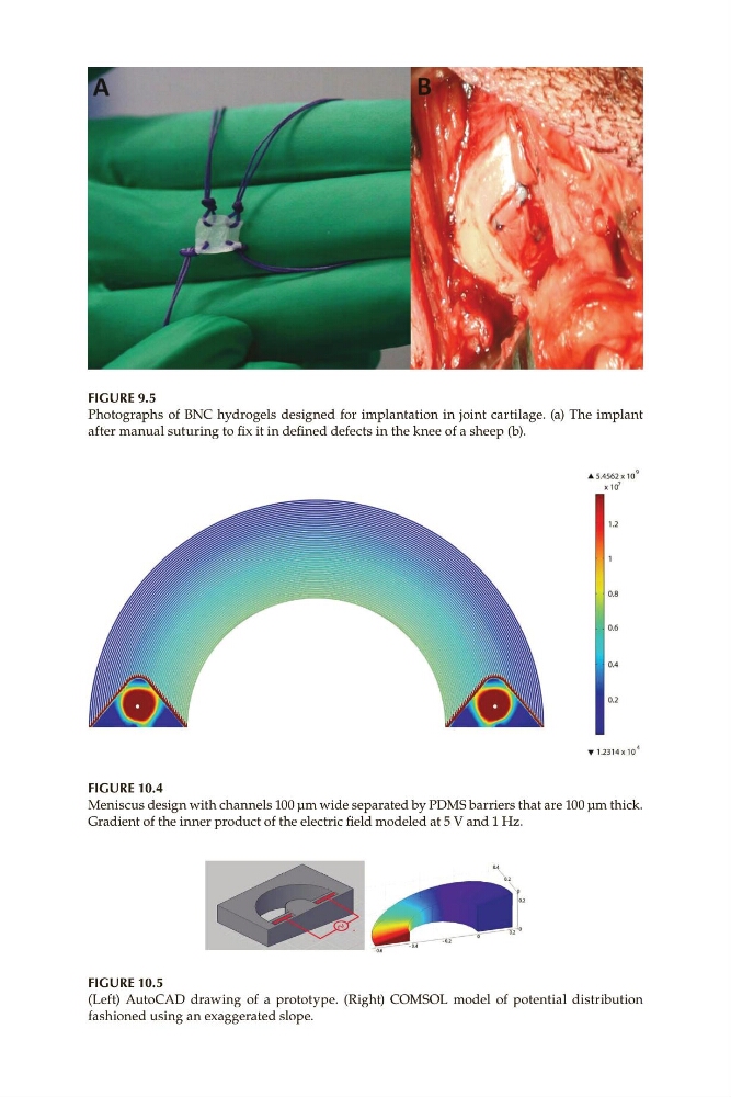
Figure 9.5
Photographs of BNC hydrogels designed for implantation in joint cartilage. (a) The implant
after manual suturing to fix it in defined defects in the knee of a sheep (b).
Figure 10.4
Meniscus design with channels 100 μm wide separated by PDMS barriers that are 100 μm thick.
Gradient of the inner product of the electric field modeled at 5 V and 1 Hz.
Figure 10.5
(Left) AutoCAD drawing of a prototype. (Right) COMSOL model of potential distribution
fashioned using an exaggerated slope.
Photographs of BNC hydrogels designed for implantation in joint cartilage. (a) The implant
after manual suturing to fix it in defined defects in the knee of a sheep (b).
Figure 10.4
Meniscus design with channels 100 μm wide separated by PDMS barriers that are 100 μm thick.
Gradient of the inner product of the electric field modeled at 5 V and 1 Hz.
Figure 10.5
(Left) AutoCAD drawing of a prototype. (Right) COMSOL model of potential distribution
fashioned using an exaggerated slope.