

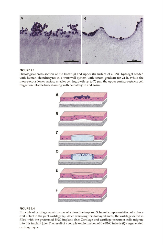
Figure 9.1
Histological cross-section of the lower (a) and upper (b) surface of a BNC hydrogel seeded
with human chondrocytes in a transwell system with serum gradient for 24 h. While the
more porous lower surface enables cell ingrowth up to 70 μm, the upper surface restricts cell
migration into the bulk staining with hematoxylin and eosin.
Figure 9.4
Principle of cartilage repair by use of a bioactive implant. Schematic representation of a chondral defect in the joint cartilage (a). After removing the damaged areas, the cartilage defect is
filled with the preformed BNC implant. (b,c) Cartilage and cartilage precursor cells migrate
into this implant (d,e). The result of a complete colonization of the BNC inlay is (f) a regenerated
cartilage layer.
Histological cross-section of the lower (a) and upper (b) surface of a BNC hydrogel seeded
with human chondrocytes in a transwell system with serum gradient for 24 h. While the
more porous lower surface enables cell ingrowth up to 70 μm, the upper surface restricts cell
migration into the bulk staining with hematoxylin and eosin.
Figure 9.4
Principle of cartilage repair by use of a bioactive implant. Schematic representation of a chondral defect in the joint cartilage (a). After removing the damaged areas, the cartilage defect is
filled with the preformed BNC implant. (b,c) Cartilage and cartilage precursor cells migrate
into this implant (d,e). The result of a complete colonization of the BNC inlay is (f) a regenerated
cartilage layer.