

251
Craniofacial Development and Disorders
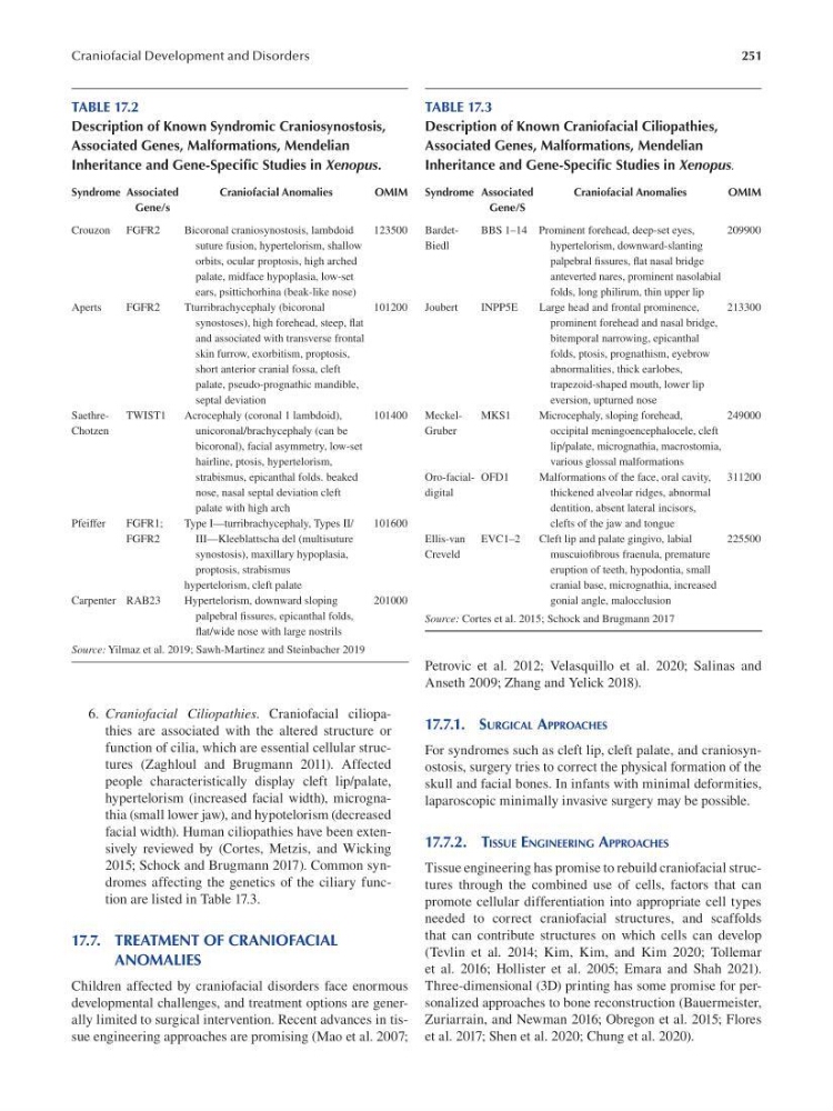
TABLE 17.2
Description of Known Syndromic Craniosynostosis,
Associated Genes, Malformations, Mendelian
Inheritance and Gene-Specific Studies in Xenopus.
Syndrome Associated
Craniofacial Anomalies
OMIM
Gene/s
Crouzon FGFR2
Bicoronal craniosynostosis, lambdoid
123500
suture fusion, hypertelorism, shallow
orbits, ocular proptosis, high arched
palate, midface hypoplasia, low-set
ears, psittichorhina (beak-like nose)
Aperts
FGFR2
Tturribrachycephaly (bicoronal
101200
synostoses), high forehead, steep, f at
and associated with transverse frontal
skin furrow, exorbitism, proptosis,
short anterior cranial fossa, cleft
palate, pseudo-prognathic mandible,
septal deviation
SaethreTWIST1
Acrocephaly (coronal 1 lambdoid),
101400
Chotzen
unicoronal/brachycephaly (can be
bicoronal), facial asymmetry, low-set
hairline, ptosis, hypertelorism,
strabismus, epicanthal folds. beaked
nose, nasal septal deviation cleft
palate with high arch
Pfeiffer
FGFR1;
Type I—turribrachycephaly, Types II/
101600
FGFR2
III—Kleeblattscha del (multisuture
synostosis), maxillary hypoplasia,
proptosis, strabismus
hypertelorism, cleft palate
Carpenter RAB23
Hypertelorism, downward sloping
201000
palpebral fssures, epicanthal folds,
fat/wide nose with large nostrils
Source: Yilmaz et al. 2019 ; Sawh-Martinez and Steinbacher 2019
6. Craniofacial Ciliopathies. Craniofacial ciliopathies are associated with the altered structure or
function of cilia, which are essential cellular structures ( Zaghloul and Brugmann 2011). Affected
people characteristically display cleft lip/palate,
hypertelorism (increased facial width), micrognathia (small lower jaw), and hypotelorism (decreased
facial width). Human ciliopathies have been extensively reviewed by (Cortes, Metzis, and Wicking
2015; Schock and Brugmann 2017). Common syndromes affecting the genetics of the ciliary function are listed in Table 17.3.
17.7. TREATMENT OF CRANIOFACIAL
ANOMALIES
Children affected by craniofacial disorders face enormous
developmental challenges, and treatment options are generally limited to surgical intervention. Recent advances in tissue engineering approaches are promising (Mao et al. 2007;
TABLE 17.3
Description of Known Craniofacial Ciliopathies,
Associated Genes, Malformations, Mendelian
Inheritance and Gene-Specific Studies in Xenopus.
Syndrome Associated
Craniofacial Anomalies
OMIM
Gene/S
BardetBBS 1–14 Prominent forehead, deep-set eyes,
209900
Biedl
hypertelorism, downward-slanting
palpebral f ssures, fat nasal bridge
anteverted nares, prominent nasolabial
folds, long philirum, thin upper lip
Joubert
INPP5E
Large head and frontal prominence,
213300
prominent forehead and nasal bridge,
bitemporal narrowing, epicanthal
folds, ptosis, prognathism, eyebrow
abnormalities, thick earlobes,
trapezoid-shaped mouth, lower lip
eversion, upturned nose
MeckelMKS1
Microcephaly, sloping forehead,
249000
Gruber
occipital meningoencephalocele, cleft
lip/palate, micrognathia, macrostomia,
various glossal malformations
Oro-facial- OFD1
Malformations of the face, oral cavity, 311200
digital
thickened alveolar ridges, abnormal
dentition, absent lateral incisors,
clefts of the jaw and tongue
Ellis-van EVC1–2
Cleft lip and palate gingivo, labial
225500
Creveld
muscuiofbrous fraenula, premature
eruption of teeth, hypodontia, small
cranial base, micrognathia, increased
gonial angle, malocclusion
Source: Cortes et al. 2015 ; Schock and Brugmann 2017
Petrovic et al. 2012; Velasquillo et al. 2020; Salinas and
Anseth 2009; Zhang and Yelick 2018).
17.7.1. SURGICAL APPROACHES
For syndromes such as cleft lip, cleft palate, and craniosynostosis, surgery tries to correct the physical formation of the
skull and facial bones. In infants with minimal deformities,
laparoscopic minimally invasive surgery may be possible.
17.7.2. TISSUE ENGINEERING APPROACHES
Tissue engineering has promise to rebuild craniofacial structures through the combined use of cells, factors that can
promote cellular differentiation into appropriate cell types
needed to correct craniofacial structures, and scaffolds
that can contribute structures on which cells can develop
(Tevlin et al. 2014; Kim, Kim, and Kim 2020; Tollemar
et al. 2016; Hollister et al. 2005; Emara and Shah 2021).
Three-dimensional (3D) printing has some promise for personalized approaches to bone reconstruction (Bauermeister,
Zuriarrain, and Newman 2016; Obregon et al. 2015; Flores
et al. 2017; Shen et al. 2020; Chung et al. 2020).