

252
Xenopus
17.7.3. CELL-BASED APPROACHES
The idea here is to use precursor cells (osteoblasts, chondrocytes, fbroblasts) to produce cartilage and bone tissue that
can repair craniofacial anomalies (Meijer et al. 2007; Teven
et al. 2012; Teven et al. 2015). Use of stem cells for craniofacial repair may be extremely useful in the future, particularly autologous cells derived from the affected person, such
as mesenchymal, adipogenic, skeletal stem cells, or induced
pluripotential stem cells ( Zuk 2008; Perez et al. 2018; Griff n
et al. 2014; Tevlin, Longaker, and Wan 2020; Borrelli et al.
2020; Velasquillo, Madrazo-Ibarra et al. 2020).
17.8. CONTRIBUTION OF XENOPUS
TO UNDERSTANDING
CRANIOFACIAL ANOMALIES
Craniofacial anomalies are prevalent and severe, and there is
an unmet need to defne the genetic or environmental perturbations that are associated with and cause these. Contributions
from Xenopus are providing insight into genes associated with
human craniofacial disorders by two approaches. The f rst is
understanding from fundamental research as to what genes
govern craniofacial development, as explored in Sections
3 and 4 of this chapter. If such genes are later identif ed in
human genome wide association screens (GWASs) as associated with a craniofacial anomaly, there will already be information available to help understand mechanisms that may
contribute to the anomaly. In an example from our group, we
identif ed zic1/opl as a gene required for neural determination and patterning (Kuo et al. 1998). Recently, heterozygous
mutations in the third exon of ZIC1 encoding the C-terminus
of the protein are associated with craniosynostosis involving
the coronal sutures (Twigg et al. 2015).
The second approach is to screen GWAS hits for their
effects on craniofacial development (Khandelwal et al. 2013;
Sanchez-Lara 2015; Yu et al. 2017; Saleem et al. 2019) and to
use Xenopus as an assay system for gene activity (Abu-Daya,
Khokha, and Zimmerman 2012; Hwang, Marquez, and Khokha
2019). This can be done by loss-of-function using antisense
techniques or CRISPR-mediated genome editing in the F0
embryo. For gene variants that appear to be gain-of-function,
RNA injection or (less successful) DNA expression constructs
injected into the embryo can be used. Tadpoles can be assayed
after the craniofacial cartilages have formed for anomalies that
may be similar to affected people. Where multiple genes are
implicated through human genetic analysis, Xenopus assays
can help sort out which are the key genes contributing to such
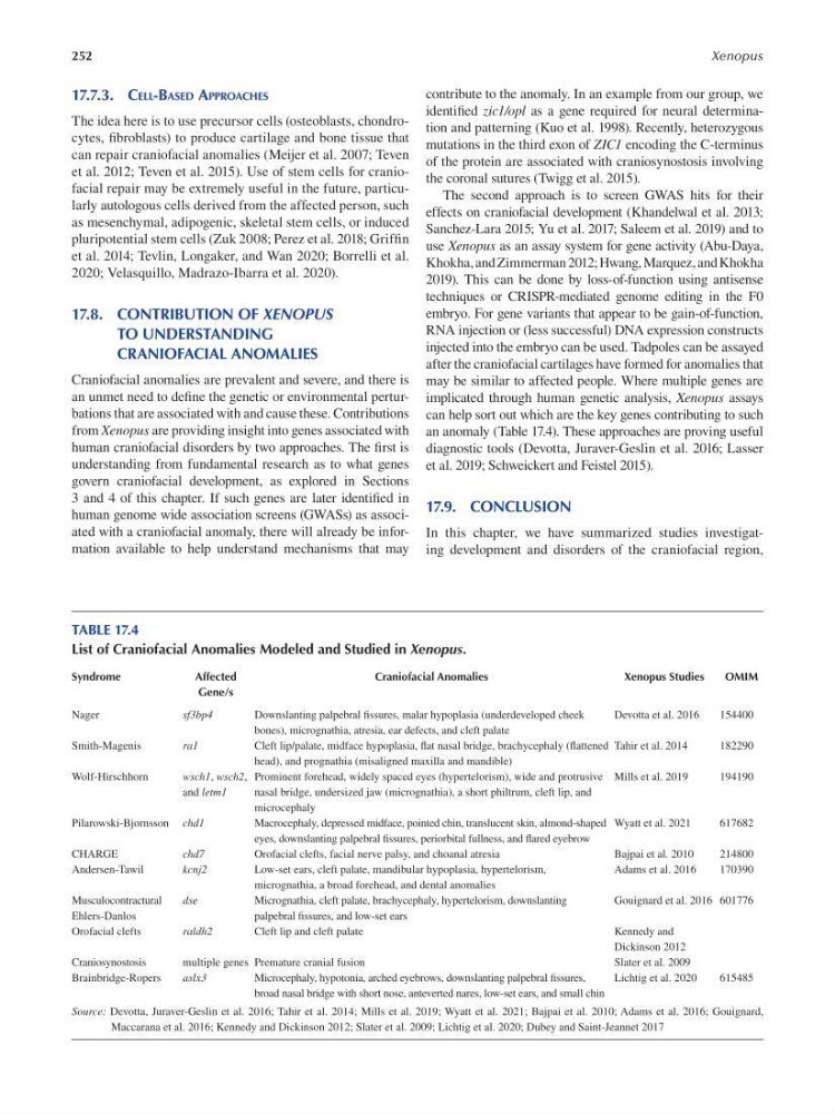
an anomaly (Table 17.4). These approaches are proving useful
diagnostic tools (Devotta, Juraver-Geslin et al. 2016; Lasser
et al. 2019; Schweickert and Feistel 2015).
17.9. CONCLUSION
In this chapter, we have summarized studies investigating development and disorders of the craniofacial region,
TABLE 17.4
List of Craniofacial Anomalies Modeled and Studied in Xenopus.
Syndrome
Affected
Craniofacial Anomalies
Xenopus Studies
OMIM
Gene/s
Nager
sf3bp4
Downslanting palpebral fssures, malar hypoplasia (underdeveloped cheek
Devotta et al. 2016
154400
bones), micrognathia, atresia, ear defects, and cleft palate
Smith-Magenis
ra1
Cleft lip/palate, midface hypoplasia, fat nasal bridge, brachycephaly (f attened Tahir et al. 2014
182290
head), and prognathia (misaligned maxilla and mandible)
Wolf-Hirschhorn
wsch1, wsch2, Prominent forehead, widely spaced eyes (hypertelorism), wide and protrusive Mills et al. 2019
194190
and letm1
nasal bridge, undersized jaw (micrognathia), a short philtrum, cleft lip, and
microcephaly
Pilarowski-Bjornsson chd1
Macrocephaly, depressed midface, pointed chin, translucent skin, almond-shaped Wyatt et al. 2021
617682
eyes, downslanting palpebral fssures, periorbital fullness, and f ared eyebrow
CHARGE
chd7
Orofacial clefts, facial nerve palsy, and choanal atresia
Bajpai et al. 2010
214800
Andersen-Tawil
kcnj2
Low-set ears, cleft palate, mandibular hypoplasia, hypertelorism,
Adams et al. 2016
170390
micrognathia, a broad forehead, and dental anomalies
Musculocontractural
dse
Micrognathia, cleft palate, brachycephaly, hypertelorism, downslanting
Gouignard et al. 2016 601776
Ehlers-Danlos
palpebral fssures, and low-set ears
Orofacial clefts
raldh2
Cleft lip and cleft palate
Kennedy and
Dickinson 2012
Craniosynostosis
multiple genes Premature cranial fusion
Slater et al. 2009
Brainbridge-Ropers
aslx3
Microcephaly, hypotonia, arched eyebrows, downslanting palpebral f ssures,
Lichtig et al. 2020
615485
broad nasal bridge with short nose, anteverted nares, low-set ears, and small chin
Source : Devotta, Juraver-Geslin et al. 2016 ; Tahir et al. 2014; Mills et al. 2019; Wyatt et al. 2021; Bajpai et al. 2010; Adams et al. 2016; Gouignard,
Maccarana et al. 2016 ; Kennedy and Dickinson 2012; Slater et al. 2009; Lichtig et al. 2020; Dubey and Saint-Jeannet 2017
Xenopus
17.7.3. CELL-BASED APPROACHES
The idea here is to use precursor cells (osteoblasts, chondrocytes, fbroblasts) to produce cartilage and bone tissue that
can repair craniofacial anomalies (Meijer et al. 2007; Teven
et al. 2012; Teven et al. 2015). Use of stem cells for craniofacial repair may be extremely useful in the future, particularly autologous cells derived from the affected person, such
as mesenchymal, adipogenic, skeletal stem cells, or induced
pluripotential stem cells ( Zuk 2008; Perez et al. 2018; Griff n
et al. 2014; Tevlin, Longaker, and Wan 2020; Borrelli et al.
2020; Velasquillo, Madrazo-Ibarra et al. 2020).
17.8. CONTRIBUTION OF XENOPUS
TO UNDERSTANDING
CRANIOFACIAL ANOMALIES
Craniofacial anomalies are prevalent and severe, and there is
an unmet need to defne the genetic or environmental perturbations that are associated with and cause these. Contributions
from Xenopus are providing insight into genes associated with
human craniofacial disorders by two approaches. The f rst is
understanding from fundamental research as to what genes
govern craniofacial development, as explored in Sections
3 and 4 of this chapter. If such genes are later identif ed in
human genome wide association screens (GWASs) as associated with a craniofacial anomaly, there will already be information available to help understand mechanisms that may
contribute to the anomaly. In an example from our group, we
identif ed zic1/opl as a gene required for neural determination and patterning (Kuo et al. 1998). Recently, heterozygous
mutations in the third exon of ZIC1 encoding the C-terminus
of the protein are associated with craniosynostosis involving
the coronal sutures (Twigg et al. 2015).
The second approach is to screen GWAS hits for their
effects on craniofacial development (Khandelwal et al. 2013;
Sanchez-Lara 2015; Yu et al. 2017; Saleem et al. 2019) and to
use Xenopus as an assay system for gene activity (Abu-Daya,
Khokha, and Zimmerman 2012; Hwang, Marquez, and Khokha
2019). This can be done by loss-of-function using antisense
techniques or CRISPR-mediated genome editing in the F0
embryo. For gene variants that appear to be gain-of-function,
RNA injection or (less successful) DNA expression constructs
injected into the embryo can be used. Tadpoles can be assayed
after the craniofacial cartilages have formed for anomalies that
may be similar to affected people. Where multiple genes are
implicated through human genetic analysis, Xenopus assays
can help sort out which are the key genes contributing to such
an anomaly (Table 17.4). These approaches are proving useful
diagnostic tools (Devotta, Juraver-Geslin et al. 2016; Lasser
et al. 2019; Schweickert and Feistel 2015).
17.9. CONCLUSION
In this chapter, we have summarized studies investigating development and disorders of the craniofacial region,
TABLE 17.4
List of Craniofacial Anomalies Modeled and Studied in Xenopus.
Syndrome
Affected
Craniofacial Anomalies
Xenopus Studies
OMIM
Gene/s
Nager
sf3bp4
Downslanting palpebral fssures, malar hypoplasia (underdeveloped cheek
Devotta et al. 2016
154400
bones), micrognathia, atresia, ear defects, and cleft palate
Smith-Magenis
ra1
Cleft lip/palate, midface hypoplasia, fat nasal bridge, brachycephaly (f attened Tahir et al. 2014
182290
head), and prognathia (misaligned maxilla and mandible)
Wolf-Hirschhorn
wsch1, wsch2, Prominent forehead, widely spaced eyes (hypertelorism), wide and protrusive Mills et al. 2019
194190
and letm1
nasal bridge, undersized jaw (micrognathia), a short philtrum, cleft lip, and
microcephaly
Pilarowski-Bjornsson chd1
Macrocephaly, depressed midface, pointed chin, translucent skin, almond-shaped Wyatt et al. 2021
617682
eyes, downslanting palpebral fssures, periorbital fullness, and f ared eyebrow
CHARGE
chd7
Orofacial clefts, facial nerve palsy, and choanal atresia
Bajpai et al. 2010
214800
Andersen-Tawil
kcnj2
Low-set ears, cleft palate, mandibular hypoplasia, hypertelorism,
Adams et al. 2016
170390
micrognathia, a broad forehead, and dental anomalies
Musculocontractural
dse
Micrognathia, cleft palate, brachycephaly, hypertelorism, downslanting
Gouignard et al. 2016 601776
Ehlers-Danlos
palpebral fssures, and low-set ears
Orofacial clefts
raldh2
Cleft lip and cleft palate
Kennedy and
Dickinson 2012
Craniosynostosis
multiple genes Premature cranial fusion
Slater et al. 2009
Brainbridge-Ropers
aslx3
Microcephaly, hypotonia, arched eyebrows, downslanting palpebral f ssures,
Lichtig et al. 2020
615485
broad nasal bridge with short nose, anteverted nares, low-set ears, and small chin
Source : Devotta, Juraver-Geslin et al. 2016 ; Tahir et al. 2014; Mills et al. 2019; Wyatt et al. 2021; Bajpai et al. 2010; Adams et al. 2016; Gouignard,
Maccarana et al. 2016 ; Kennedy and Dickinson 2012; Slater et al. 2009; Lichtig et al. 2020; Dubey and Saint-Jeannet 2017