

250
Xenopus
during pregnancy have been implicated in craniofacial anomalies.
3. Vitamin def ciency. A maternal diet lacking Vitamin
A, Vitamin D, folic acid (Vitamin B9), or Vitamin
B12 is associated with higher risk of CFAs such as
cleft lip and cleft palate (Maldonado et al. 2021;
Wahl et al. 2015; Finkelstein, Layden, and Stover
2015; Pannia et al. 2016; Mulligan et al. 2010;
Clagett-Dame and Knutson 2011).
17.6.2. CLASSES OF CRANIOFACIAL ANOMALIES
1. Cleft lip or cleft palate. Cleft lip and cleft palate are
the most common congenital craniofacial anomalies seen at birth. In cleft lip, the lip does not form
properly. The degree of the cleft lip varies greatly,
from a mild notch to a severe large opening up
to the base of the nose. In cleft palate, the palatal
shelves do not close completely, leaving an opening extending into the nasal cavity. The cleft can
extend from the front of the mouth to the throat and
may also include the lip.
2. Craniofacial microsomia. In these cases, one side
of the face is smaller than the other, giving rise to
facial asymmetry. Typically, only the lower part of
the face is affected, with a fat cheek due to anomalous bone growth and an underdeveloped jaw (maxillary or mandibular hypoplasia). The external ears
are malformed or absent. Craniofacial microsomia
with maxillary or mandibular hypoplasia may be
referred to as hemifacial microsomia and includes
Goldenhar syndrome, branchial arch syndrome,
and lateral facial dysplasia (Birgfeld and Heike
2012; Bogusiak, Puch, and Arkuszewski 2017;
Monahan et al. 2001; Brandstetter and Patel 2016).
3. Neurocristopathies. Neurocristopathies are perturbations in the formation, migration, and/or differentiation of neural crest cells (Watt and Trainor
2014; Vega-Lopez et al. 2018). Cranial neural crest
cells migrate into the frst and second pharyngeal
arches of a developing embryo. In humans, the
cells in the frst pharyngeal arch give rise to the
maxilla, zygoma, palate, mandible, malleus, incus,
muscle of mastication, and parts of the trigeminal
sensory ganglion. Those of the second pharyngeal
arch give rise to the hyoid cartilage, stapes, facial
muscles, and parts of the facial sensory ganglia
(Johnson et al. 2011; Richany, Bast, and Anson
1955; Bast, Anson, and Richany 1955). Disruptions
to the frst and second pharyngeal arches are associated with craniofacial anomalies known as facial
dysostoses (Trainor and Andrews 2013). These
include Treacher Collins syndrome, Nager syndrome, Miller syndrome, and Goldenhar syndrome
(Trainor and Andrews 2013; Sato et al. 2019). The
vast amount of data on neural crest development
in Xenopus provide an important model system
in which to study these syndromes (Table 17.4)
(Gouignard et al. 2016; Schwenty-Lara, Pauli, and
Borchers 2020; Devotta et al. 2016).
4. Craniofacial Dysmorphism. Craniofacial dysmorphism is defned as an abnormally formed
craniofacial structure. This can include brachycephaly (fattened back of the head), highly arched
eyebrows, monobrow, low-set ears, microdontism
(smaller teeth), and generalized gingival hyperplasia (overgrowth of gums around the teeth). Some of
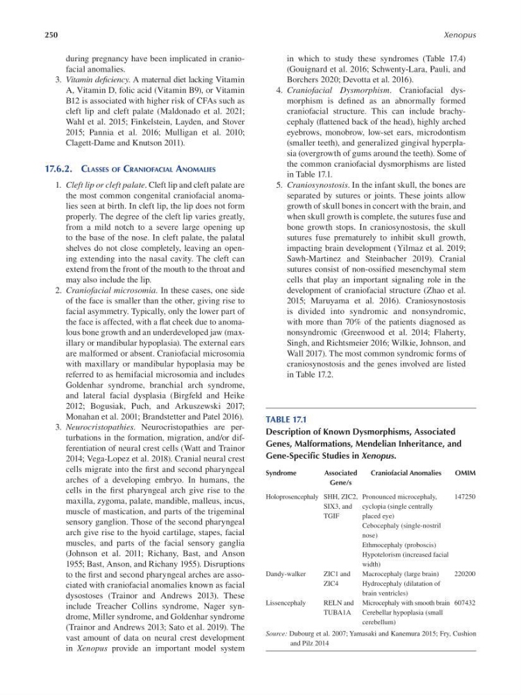
the common craniofacial dysmorphisms are listed
in Table 17.1.
5. Craniosynostosis. In the infant skull, the bones are
separated by sutures or joints. These joints allow
growth of skull bones in concert with the brain, and
when skull growth is complete, the sutures fuse and
bone growth stops. In craniosynostosis, the skull
sutures fuse prematurely to inhibit skull growth,
impacting brain development (Yilmaz et al. 2019;
Sawh-Martinez and Steinbacher 2019). Cranial
sutures consist of non-ossifed mesenchymal stem
cells that play an important signaling role in the
development of craniofacial structure ( Zhao et al.
2015; Maruyama et al. 2016). Craniosynostosis
is divided into syndromic and nonsyndromic,
with more than 70% of the patients diagnosed as
nonsyndromic (Greenwood et al. 2014; Flaherty,
Singh, and Richtsmeier 2016; Wilkie, Johnson, and
Wall 2017). The most common syndromic forms of
craniosynostosis and the genes involved are listed
in Table 17.2.
TABLE 17.1
Description of Known Dysmorphisms, Associated
Genes, Malformations, Mendelian Inheritance, and
Gene-Specific Studies in Xenopus.
Syndrome
Associated
Craniofacial Anomalies
OMIM
Gene/s
Holoprosencephaly SHH, ZIC2, Pronounced microcephaly,
147250
SIX3, and cyclopia (single centrally
TGIF
placed eye)
Cebocephaly (single-nostril
nose)
Ethmocephaly (proboscis)
Hypotelorism (increased facial
width)
Dandy-walker
ZIC1 and
Macrocephaly (large brain)
220200
ZIC4
Hydrocephaly (dilatation of
brain ventricles)
Lissencephaly
RELN and Microcephaly with smooth brain 607432
TUBA1A
Cerebellar hypoplasia (small
cerebellum)
Source: Dubourg et al. 2007; Yamasaki and Kanemura 2015; Fry, Cushion
and Pilz 2014
Xenopus
during pregnancy have been implicated in craniofacial anomalies.
3. Vitamin def ciency. A maternal diet lacking Vitamin
A, Vitamin D, folic acid (Vitamin B9), or Vitamin
B12 is associated with higher risk of CFAs such as
cleft lip and cleft palate (Maldonado et al. 2021;
Wahl et al. 2015; Finkelstein, Layden, and Stover
2015; Pannia et al. 2016; Mulligan et al. 2010;
Clagett-Dame and Knutson 2011).
17.6.2. CLASSES OF CRANIOFACIAL ANOMALIES
1. Cleft lip or cleft palate. Cleft lip and cleft palate are
the most common congenital craniofacial anomalies seen at birth. In cleft lip, the lip does not form
properly. The degree of the cleft lip varies greatly,
from a mild notch to a severe large opening up
to the base of the nose. In cleft palate, the palatal
shelves do not close completely, leaving an opening extending into the nasal cavity. The cleft can
extend from the front of the mouth to the throat and
may also include the lip.
2. Craniofacial microsomia. In these cases, one side
of the face is smaller than the other, giving rise to
facial asymmetry. Typically, only the lower part of
the face is affected, with a fat cheek due to anomalous bone growth and an underdeveloped jaw (maxillary or mandibular hypoplasia). The external ears
are malformed or absent. Craniofacial microsomia
with maxillary or mandibular hypoplasia may be
referred to as hemifacial microsomia and includes
Goldenhar syndrome, branchial arch syndrome,
and lateral facial dysplasia (Birgfeld and Heike
2012; Bogusiak, Puch, and Arkuszewski 2017;
Monahan et al. 2001; Brandstetter and Patel 2016).
3. Neurocristopathies. Neurocristopathies are perturbations in the formation, migration, and/or differentiation of neural crest cells (Watt and Trainor
2014; Vega-Lopez et al. 2018). Cranial neural crest
cells migrate into the frst and second pharyngeal
arches of a developing embryo. In humans, the
cells in the frst pharyngeal arch give rise to the
maxilla, zygoma, palate, mandible, malleus, incus,
muscle of mastication, and parts of the trigeminal
sensory ganglion. Those of the second pharyngeal
arch give rise to the hyoid cartilage, stapes, facial
muscles, and parts of the facial sensory ganglia
(Johnson et al. 2011; Richany, Bast, and Anson
1955; Bast, Anson, and Richany 1955). Disruptions
to the frst and second pharyngeal arches are associated with craniofacial anomalies known as facial
dysostoses (Trainor and Andrews 2013). These
include Treacher Collins syndrome, Nager syndrome, Miller syndrome, and Goldenhar syndrome
(Trainor and Andrews 2013; Sato et al. 2019). The
vast amount of data on neural crest development
in Xenopus provide an important model system
in which to study these syndromes (Table 17.4)
(Gouignard et al. 2016; Schwenty-Lara, Pauli, and
Borchers 2020; Devotta et al. 2016).
4. Craniofacial Dysmorphism. Craniofacial dysmorphism is defned as an abnormally formed
craniofacial structure. This can include brachycephaly (fattened back of the head), highly arched
eyebrows, monobrow, low-set ears, microdontism
(smaller teeth), and generalized gingival hyperplasia (overgrowth of gums around the teeth). Some of
the common craniofacial dysmorphisms are listed
in Table 17.1.
5. Craniosynostosis. In the infant skull, the bones are
separated by sutures or joints. These joints allow
growth of skull bones in concert with the brain, and
when skull growth is complete, the sutures fuse and
bone growth stops. In craniosynostosis, the skull
sutures fuse prematurely to inhibit skull growth,
impacting brain development (Yilmaz et al. 2019;
Sawh-Martinez and Steinbacher 2019). Cranial
sutures consist of non-ossifed mesenchymal stem
cells that play an important signaling role in the
development of craniofacial structure ( Zhao et al.
2015; Maruyama et al. 2016). Craniosynostosis
is divided into syndromic and nonsyndromic,
with more than 70% of the patients diagnosed as
nonsyndromic (Greenwood et al. 2014; Flaherty,
Singh, and Richtsmeier 2016; Wilkie, Johnson, and
Wall 2017). The most common syndromic forms of
craniosynostosis and the genes involved are listed
in Table 17.2.
TABLE 17.1
Description of Known Dysmorphisms, Associated
Genes, Malformations, Mendelian Inheritance, and
Gene-Specific Studies in Xenopus.
Syndrome
Associated
Craniofacial Anomalies
OMIM
Gene/s
Holoprosencephaly SHH, ZIC2, Pronounced microcephaly,
147250
SIX3, and cyclopia (single centrally
TGIF
placed eye)
Cebocephaly (single-nostril
nose)
Ethmocephaly (proboscis)
Hypotelorism (increased facial
width)
Dandy-walker
ZIC1 and
Macrocephaly (large brain)
220200
ZIC4
Hydrocephaly (dilatation of
brain ventricles)
Lissencephaly
RELN and Microcephaly with smooth brain 607432
TUBA1A
Cerebellar hypoplasia (small
cerebellum)
Source: Dubourg et al. 2007; Yamasaki and Kanemura 2015; Fry, Cushion
and Pilz 2014