

methylenes and suggested the presence of one unit of lactobacillic acid.
A cosy experiment allowed to assign protons H-1, H-2 and H-3 corresponding to the glycerol moiety and showed that the third methylene (δ 4.06 ppm) was coupled only to another one located at δ 3.14ppm,
suggesting the presence of a phosphatidylethanolamine moiety in
agreement with the chemical shift values (Kohama et al., 1991; Batrakov & Nikitin, 1996). A 3I P NMR 121 MHz experiment was carried
out and allowed to observe a signal at ô - 0.6 ppm which confirmed
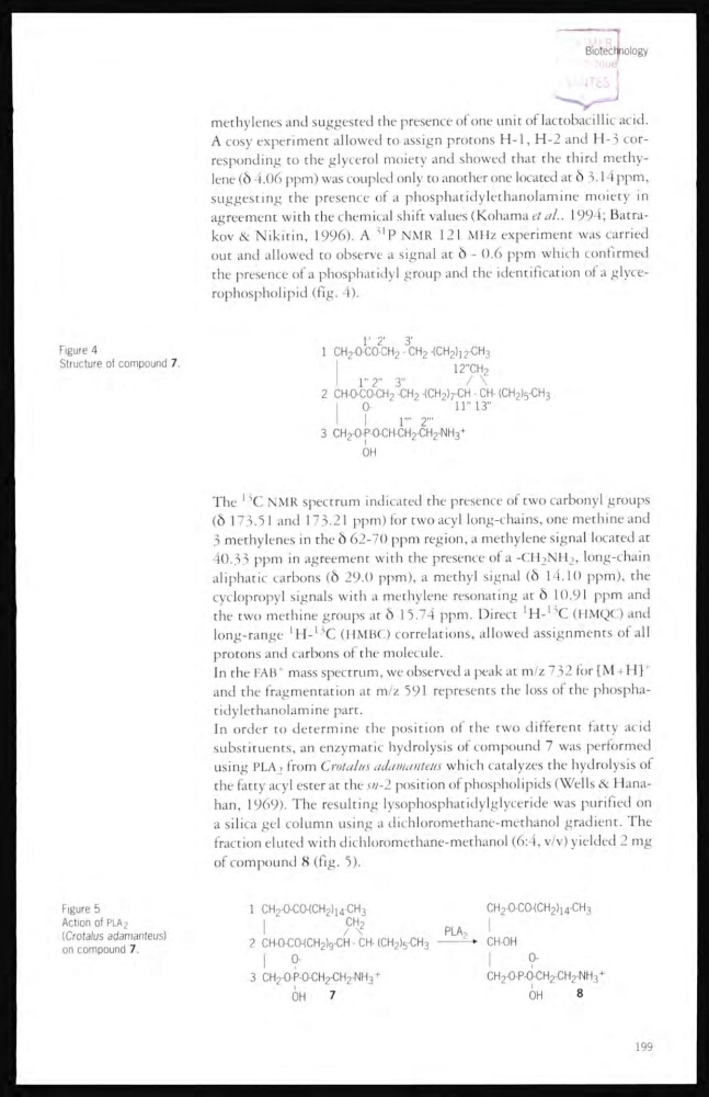
the presence of a phosphatidyl group and the identification of a glycerophospholipid (fig. 4).
Figure 4
Structure of compound 7
The
13 C NMR spectrum indicated the presence of two carbonyl groups
(δ 173.51 and 173.21 ppm) for two acyl long-chains, one methine and
3 methylenes in the δ 62-70 ppm region, a methylene signal located at
40.33 ppm in agreement with the presence of a -CH
2
NH
2
, long-chain
aliphatic carbons (δ 29-0 ppm), a methyl signal (ô 14.10 ppm), the
cyclopropyl signals with a methylene resonating at ô 10.91 ppm and
the two methine groups at δ 15.74 ppm. Direct 1 H- 13 C (HMQC) and
long-range
1 H15 C (HMBC) correlations, allowed assignments of all
protons and carbons of the molecule.
In the FAB
+ mass spectrum, we observed a peak at m/z 732 for [M + H] +
and the fragmentation at m/z 591 represents the loss of the phosphatidylethanolamine part.
In order to determine the position of the two different fatty acid
substituents, an enzymatic hydrolysis of compound 7 was performed
using PLA 2 from Crotalus adamanteus which catalyzes the hydrolysis of
the fatty acyl ester at the sn-2 position of phospholipids (Wells & Hanahan, 1969). The resulting lysophosphatidylglyceride was purified on
a silica gel column using a dichloromethane-methanol gradient. The
fraction eluted with dichloromethane-methanol (6:4, v/v) yielded 2 mg
of compound 8 (fig. 5).
Figure 5
Action of PLA2
(Crotalus adamanteus)
on compound 7.
199
A cosy experiment allowed to assign protons H-1, H-2 and H-3 corresponding to the glycerol moiety and showed that the third methylene (δ 4.06 ppm) was coupled only to another one located at δ 3.14ppm,
suggesting the presence of a phosphatidylethanolamine moiety in
agreement with the chemical shift values (Kohama et al., 1991; Batrakov & Nikitin, 1996). A 3I P NMR 121 MHz experiment was carried
out and allowed to observe a signal at ô - 0.6 ppm which confirmed
the presence of a phosphatidyl group and the identification of a glycerophospholipid (fig. 4).
Figure 4
Structure of compound 7
The
13 C NMR spectrum indicated the presence of two carbonyl groups
(δ 173.51 and 173.21 ppm) for two acyl long-chains, one methine and
3 methylenes in the δ 62-70 ppm region, a methylene signal located at
40.33 ppm in agreement with the presence of a -CH
2
NH
2
, long-chain
aliphatic carbons (δ 29-0 ppm), a methyl signal (ô 14.10 ppm), the
cyclopropyl signals with a methylene resonating at ô 10.91 ppm and
the two methine groups at δ 15.74 ppm. Direct 1 H- 13 C (HMQC) and
long-range
1 H15 C (HMBC) correlations, allowed assignments of all
protons and carbons of the molecule.
In the FAB
+ mass spectrum, we observed a peak at m/z 732 for [M + H] +
and the fragmentation at m/z 591 represents the loss of the phosphatidylethanolamine part.
In order to determine the position of the two different fatty acid
substituents, an enzymatic hydrolysis of compound 7 was performed
using PLA 2 from Crotalus adamanteus which catalyzes the hydrolysis of
the fatty acyl ester at the sn-2 position of phospholipids (Wells & Hanahan, 1969). The resulting lysophosphatidylglyceride was purified on
a silica gel column using a dichloromethane-methanol gradient. The
fraction eluted with dichloromethane-methanol (6:4, v/v) yielded 2 mg
of compound 8 (fig. 5).
Figure 5
Action of PLA2
(Crotalus adamanteus)
on compound 7.
199