

10. ELECTRIC ORGANS
469
2 msec
1 0 msec
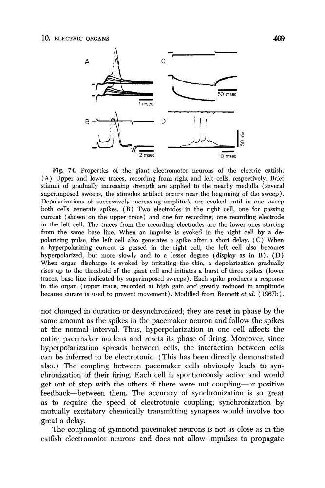
Fig. 74. Properties of the giant electromotor neurons of the electric cadish.
( A ) Upper and lower traces, recording from right and left cells, respectively. Brief
stimuli of gradually increasing strength are applied to the nearby medulla (several
superimposed sweeps, the stimulus artifact occurs near the beginning of the sweep).
Depolarizations of successively increasing amplitude are evoked until in one sweep
both cells generate spikes. ( B ) Two electrodes in the right cell, one for passing
current (shown on the upper trace) and one for recording; one recording electrode
in the left cell. The traces from the recording electrodes are the lower ones starting
from the same base line. When an impulse is evoked in the right cell by a depolarizing pulse, the left cell also generates a spike after a short delay. ( C ) When
a hyperpolarizing current is passed in the right cell, the left cell also becomes
hyperpolarized, but more slowly and to a lesser degree (display as in B ) . ( D )
When organ discharge is evoked by irritating the skin, a depolarization gradually
rises up to the threshold of the giant cell and initiates a burst of three spikes (lower
traces, base line indicated by superimposed sweeps ). Each spike produces a response
in the organ (upper trace, recorded at high gain and greatly reduced in amplitude
because curare is used to prevent movement). Modified from Bennett et al. ( 1967b).
not changed in duration or desynchronized; they are reset in phase by the
same amount as the spikes in the pacemaker neuron and follow the spikes
at the normal interval. Thus, hyperpolarization in one cell affects the
entire pacemaker nucleus and resets its phase of firing. Moreover, since
hyperpolarization spreads between cells, the interaction between cells
can be inferred to be electrotonic. (This has been directly demonstrated
also.) The coupling between pacemaker cells obviously leads to synchronization of their firing. Each cell is spontaneously active and would
get out of step with the others if there were not coupling-or positive
feedback-between them. The accuracy of synchronization is so great
as to require the speed of electrotonic coupling; synchronization by
mutually excitatory chemically transmitting synapses would involve too
great a delay.
The coupling of gymnotid pacemaker neurons is not as close as in the
catfish electromotor neurons and does not allow impulses to propagate
469
2 msec
1 0 msec
Fig. 74. Properties of the giant electromotor neurons of the electric cadish.
( A ) Upper and lower traces, recording from right and left cells, respectively. Brief
stimuli of gradually increasing strength are applied to the nearby medulla (several
superimposed sweeps, the stimulus artifact occurs near the beginning of the sweep).
Depolarizations of successively increasing amplitude are evoked until in one sweep
both cells generate spikes. ( B ) Two electrodes in the right cell, one for passing
current (shown on the upper trace) and one for recording; one recording electrode
in the left cell. The traces from the recording electrodes are the lower ones starting
from the same base line. When an impulse is evoked in the right cell by a depolarizing pulse, the left cell also generates a spike after a short delay. ( C ) When
a hyperpolarizing current is passed in the right cell, the left cell also becomes
hyperpolarized, but more slowly and to a lesser degree (display as in B ) . ( D )
When organ discharge is evoked by irritating the skin, a depolarization gradually
rises up to the threshold of the giant cell and initiates a burst of three spikes (lower
traces, base line indicated by superimposed sweeps ). Each spike produces a response
in the organ (upper trace, recorded at high gain and greatly reduced in amplitude
because curare is used to prevent movement). Modified from Bennett et al. ( 1967b).
not changed in duration or desynchronized; they are reset in phase by the
same amount as the spikes in the pacemaker neuron and follow the spikes
at the normal interval. Thus, hyperpolarization in one cell affects the
entire pacemaker nucleus and resets its phase of firing. Moreover, since
hyperpolarization spreads between cells, the interaction between cells
can be inferred to be electrotonic. (This has been directly demonstrated
also.) The coupling between pacemaker cells obviously leads to synchronization of their firing. Each cell is spontaneously active and would
get out of step with the others if there were not coupling-or positive
feedback-between them. The accuracy of synchronization is so great
as to require the speed of electrotonic coupling; synchronization by
mutually excitatory chemically transmitting synapses would involve too
great a delay.
The coupling of gymnotid pacemaker neurons is not as close as in the
catfish electromotor neurons and does not allow impulses to propagate