

426
M. V. L. BENNETI
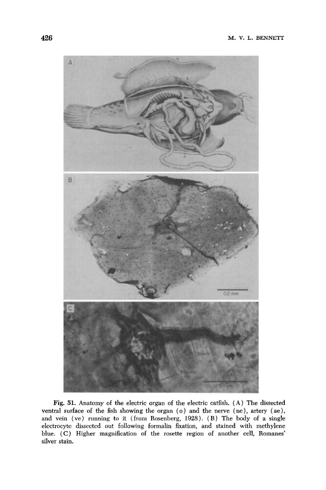
Fig. 51. Anatomy of the electric organ of the electric catfish. ( A ) The dissected
ventral surface of the fish showing the organ ( 0 ) and the nerve ( n e ) , artery ( a e ) ,
and vein (ve) running to it (from Rosenberg, 1928). ( B ) The body of a single
electrocyte dissected out following forrnalin fixation, and stained with methylene
blue. ( C ) Higher magnification of the rosette region of another cell, Romanes’
silver stain.
M. V. L. BENNETI
Fig. 51. Anatomy of the electric organ of the electric catfish. ( A ) The dissected
ventral surface of the fish showing the organ ( 0 ) and the nerve ( n e ) , artery ( a e ) ,
and vein (ve) running to it (from Rosenberg, 1928). ( B ) The body of a single
electrocyte dissected out following forrnalin fixation, and stained with methylene
blue. ( C ) Higher magnification of the rosette region of another cell, Romanes’
silver stain.