

10. ELECTRIC ORGANS
413
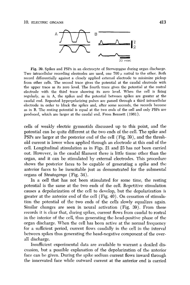
Fig. 39. Spikes and PSPs in an electrocyte of Sternopygus during organ discharge.
Two intracellular recording electrodes are used, one 700 p rostral to the other. Both
record differentially against a closely applied external electrode to minimize pickup
from other cells. The second trace gives the potential at the caudal electrode with
the upper trace as its zero level. The fourth trace gives the potential at the rostral
electrode with the third trace showing its zero level. When the cell is firing
regularly, as in A, the spikes and the potential between spikes are greater at the
caudal end. Repeated hyperpolarizing pulses are passed through a third intracellular
electrode in order to block the spikes and, after some seconds, the records become
as in B. The resting potential is equal at the two ends of the cell and only PSPs are
produced, which are larger at the caudal end. From Bennett (1961).
cells of weakly electric gymnotids discussed up to this point, and the
potential can be quite different at the two ends of the cell. The spike and
PSPs are larger at the posterior end of the cell (Fig. 39), and the threshold current is lower when applied through an electrode at this end of the
cell. Longitudinal stimulation as in Figs. 21 and 25 has not been carried
out. However, in the caudal filament there is little tissue other than the
organ, and it can be stimulated by external electrodes. This procedure
shows the posterior faces to be capable of generating a spike and the
anterior faces to be inexcitable just as demonstrated for the submental
organs of Steatogenys (Fig, 34).
In a cell that has not been stimulated for some time, the resting
potential is the same at the two ends of the cell. Repetitive stimulation
causes a depolarization of the cell to develop, but the depolarization is
greater at the anterior end of the cell (Fig. 40). On cessation of stimulation the potential of the two ends of the cells slowly equalizes again.
Similar changes are seen in neural activation (Fig. 39). From these
records it is clear that, during spikes, current flows from caudal to rostral
in the interior of the cell, thus generating the head-positive phase of the
organ discharge. When the cell has been active at the normal frequency
for a sufficient period, current flows caudally in the cell in the interval
between spikes thus generating the head-negative component of the overall discharge.
Insufficient experimental data are available to warrant a deailed discussion, but a possible explanation of the depolarization of the anterior
face can be given. During the spike sodium current flows inward through
the innervated face while outward current at the anterior end is carried
413
Fig. 39. Spikes and PSPs in an electrocyte of Sternopygus during organ discharge.
Two intracellular recording electrodes are used, one 700 p rostral to the other. Both
record differentially against a closely applied external electrode to minimize pickup
from other cells. The second trace gives the potential at the caudal electrode with
the upper trace as its zero level. The fourth trace gives the potential at the rostral
electrode with the third trace showing its zero level. When the cell is firing
regularly, as in A, the spikes and the potential between spikes are greater at the
caudal end. Repeated hyperpolarizing pulses are passed through a third intracellular
electrode in order to block the spikes and, after some seconds, the records become
as in B. The resting potential is equal at the two ends of the cell and only PSPs are
produced, which are larger at the caudal end. From Bennett (1961).
cells of weakly electric gymnotids discussed up to this point, and the
potential can be quite different at the two ends of the cell. The spike and
PSPs are larger at the posterior end of the cell (Fig. 39), and the threshold current is lower when applied through an electrode at this end of the
cell. Longitudinal stimulation as in Figs. 21 and 25 has not been carried
out. However, in the caudal filament there is little tissue other than the
organ, and it can be stimulated by external electrodes. This procedure
shows the posterior faces to be capable of generating a spike and the
anterior faces to be inexcitable just as demonstrated for the submental
organs of Steatogenys (Fig, 34).
In a cell that has not been stimulated for some time, the resting
potential is the same at the two ends of the cell. Repetitive stimulation
causes a depolarization of the cell to develop, but the depolarization is
greater at the anterior end of the cell (Fig. 40). On cessation of stimulation the potential of the two ends of the cells slowly equalizes again.
Similar changes are seen in neural activation (Fig. 39). From these
records it is clear that, during spikes, current flows from caudal to rostral
in the interior of the cell, thus generating the head-positive phase of the
organ discharge. When the cell has been active at the normal frequency
for a sufficient period, current flows caudally in the cell in the interval
between spikes thus generating the head-negative component of the overall discharge.
Insufficient experimental data are available to warrant a deailed discussion, but a possible explanation of the depolarization of the anterior
face can be given. During the spike sodium current flows inward through
the innervated face while outward current at the anterior end is carried