

412
M. V. L. BENNETT
it from reaching the spinal neurons. The stimulus however has no
noticeable effect on the following pulse indicating that the organ is
controlled rostral to the spinal cord and that the antidromic volley does
not invade the controlling nucleus. [There is actually a very slight advance of subsequent pulses ( see Bennett et d., 1967c) .]
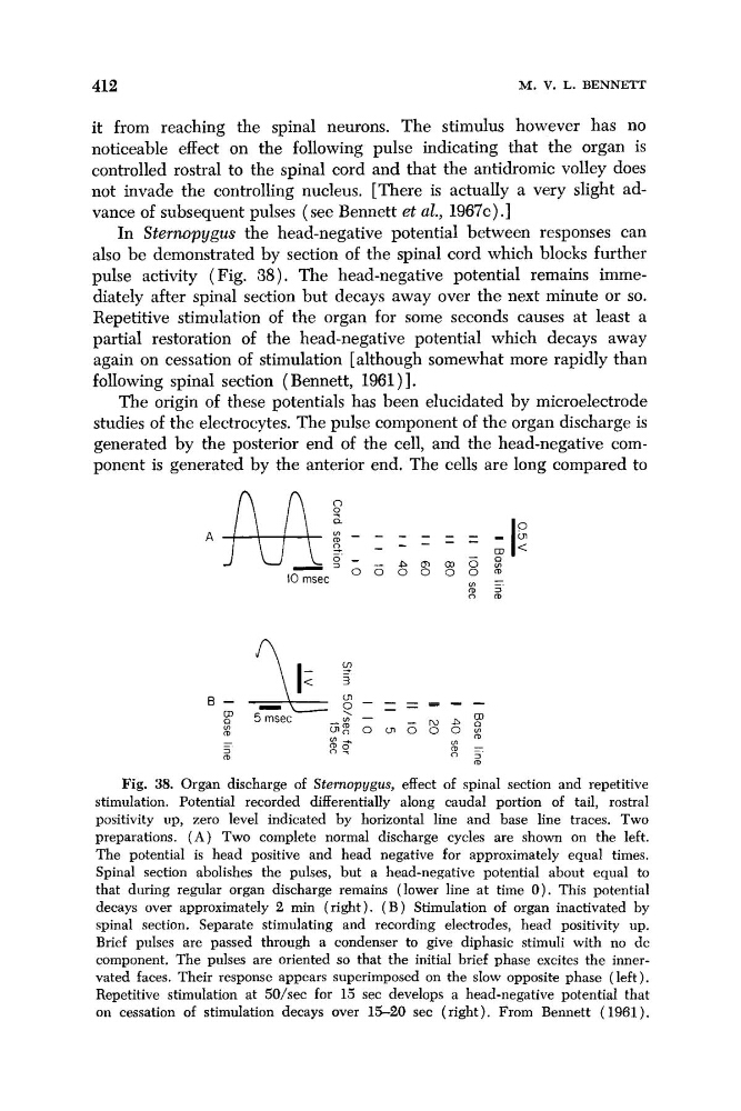
In Sterrwpygus the head-negative potential between responses can
also be demonstrated by section of the spinal cord which blocks further
pulse activity ( Fig. 38). The head-negative potential remains immediately after spinal section but decays away over the next minute or so.
Repetitive stimulation of the organ for some seconds causes at least a
partial restoration of the head-negative potential which decays away
again on cessation of stimulation [although somewhat more rapidly than
following spinal section (Bennett, 196l)I.
The origin of these potentials has been elucidated by microelectrode
studies of the electrocytes. The pulse component of the organ discharge is
generated by the posterior end of the cell, and the head-negative component is generated by the anterior end. The cells are long compared to
Fig. 38. Organ discharge of Sternopygus, effect of spinal section and repetitive
stimulation. Potential recorded differentially along caudal portion of tail, rostral
positivity up, zero level indicated by horizontal line and base line traces. Two
preparations. ( A ) Two complete normal discharge cycles are shown on the left.
The potential is head positive and head negative for approximately equal times.
Spinal section abolishes the pulses, but a head-negative potential about equal to
that during regular organ discharge remains (lower line at time 0). This potential
decays over approximately 2 min (right). ( B ) Stimulation of organ inactivated by
spinal section. Separate stimulating and recording electrodes, head positivity up.
Brief pulses are passed through a condenser to give diphasic stimuli with no dc
component. The pulses are oriented so that the initial brief phase excites the innervated faces. Their response appears superimposed on the slow opposite phase ( left ).
Repetitive stimulation at 50/sec for 15 sec develops a head-negative potential that
on cessation of stimulation decays over 15-20 sec (right). From Bennett (1961).
M. V. L. BENNETT
it from reaching the spinal neurons. The stimulus however has no
noticeable effect on the following pulse indicating that the organ is
controlled rostral to the spinal cord and that the antidromic volley does
not invade the controlling nucleus. [There is actually a very slight advance of subsequent pulses ( see Bennett et d., 1967c) .]
In Sterrwpygus the head-negative potential between responses can
also be demonstrated by section of the spinal cord which blocks further
pulse activity ( Fig. 38). The head-negative potential remains immediately after spinal section but decays away over the next minute or so.
Repetitive stimulation of the organ for some seconds causes at least a
partial restoration of the head-negative potential which decays away
again on cessation of stimulation [although somewhat more rapidly than
following spinal section (Bennett, 196l)I.
The origin of these potentials has been elucidated by microelectrode
studies of the electrocytes. The pulse component of the organ discharge is
generated by the posterior end of the cell, and the head-negative component is generated by the anterior end. The cells are long compared to
Fig. 38. Organ discharge of Sternopygus, effect of spinal section and repetitive
stimulation. Potential recorded differentially along caudal portion of tail, rostral
positivity up, zero level indicated by horizontal line and base line traces. Two
preparations. ( A ) Two complete normal discharge cycles are shown on the left.
The potential is head positive and head negative for approximately equal times.
Spinal section abolishes the pulses, but a head-negative potential about equal to
that during regular organ discharge remains (lower line at time 0). This potential
decays over approximately 2 min (right). ( B ) Stimulation of organ inactivated by
spinal section. Separate stimulating and recording electrodes, head positivity up.
Brief pulses are passed through a condenser to give diphasic stimuli with no dc
component. The pulses are oriented so that the initial brief phase excites the innervated faces. Their response appears superimposed on the slow opposite phase ( left ).
Repetitive stimulation at 50/sec for 15 sec develops a head-negative potential that
on cessation of stimulation decays over 15-20 sec (right). From Bennett (1961).