

10. ELECTRIC ORGANS
411
and there is no specialized region rostrally. The electrocytes are tubular
to spindle-shaped with the long axis parallel to the anteroposterior axis
of the fish. The cells are 1-2 mm long and 0.2 mm in diameter in a
Sternopygus 15 cm long and somewhat larger in an Eigenmunnia of
similar size (Fig. 36). They are innervated on their posterior faces. There
are about 6 columns on each side in Eigenmunnia and about 10 in
Sternopygus. The cells are accurately aligned in columns in Eigenmannia,
but they are separated by only small spaces; the amount of extracellular
space between cells is much smaller than the cells themselves. The alignment is less regular in Sternopygus, but the amount of extracellular space
is similarly small.
It may not be obvious from records such as those of Fig. 3C that the
discharge consists of head-positive pulses superimposed on a headnegative base line, and the distinction between pulse and interpulse
interval may be even less obvious in the faster firing Eigenmunniu. The
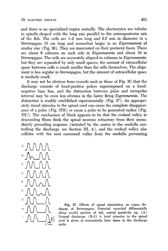
distinction is readily established experimentally (Fig. 37). An appropriately timed stimulus to the spinal cord can cause the complete disappearance of a pulse ( Fig. 37H) or cause a pulse to be generated earlier (Fig.
37C). The mechanism of block appears to be that the evoked volley in
descending fibers finds the spinal neurons refractory from their immediately preceding response (initiated by the center in the medulla controlling the discharge, see Section 111, A), and the evoked volley also
collides with the next command volley from the medulla preventing
Fig, 37. Effects of spinal stimulation on organ discharge of Sternopygus. Potential recorded differentially
along caudal portion of tail, rostra1 positivity up. ( A )
Normal discharge. ( E I ) A brief stimulus to the spinal
.fuU-i
i j l J U l 0
- o cord is given at successively later times in the discharge
10 msec
cycle.
411
and there is no specialized region rostrally. The electrocytes are tubular
to spindle-shaped with the long axis parallel to the anteroposterior axis
of the fish. The cells are 1-2 mm long and 0.2 mm in diameter in a
Sternopygus 15 cm long and somewhat larger in an Eigenmunnia of
similar size (Fig. 36). They are innervated on their posterior faces. There
are about 6 columns on each side in Eigenmunnia and about 10 in
Sternopygus. The cells are accurately aligned in columns in Eigenmannia,
but they are separated by only small spaces; the amount of extracellular
space between cells is much smaller than the cells themselves. The alignment is less regular in Sternopygus, but the amount of extracellular space
is similarly small.
It may not be obvious from records such as those of Fig. 3C that the
discharge consists of head-positive pulses superimposed on a headnegative base line, and the distinction between pulse and interpulse
interval may be even less obvious in the faster firing Eigenmunniu. The
distinction is readily established experimentally (Fig. 37). An appropriately timed stimulus to the spinal cord can cause the complete disappearance of a pulse ( Fig. 37H) or cause a pulse to be generated earlier (Fig.
37C). The mechanism of block appears to be that the evoked volley in
descending fibers finds the spinal neurons refractory from their immediately preceding response (initiated by the center in the medulla controlling the discharge, see Section 111, A), and the evoked volley also
collides with the next command volley from the medulla preventing
Fig, 37. Effects of spinal stimulation on organ discharge of Sternopygus. Potential recorded differentially
along caudal portion of tail, rostra1 positivity up. ( A )
Normal discharge. ( E I ) A brief stimulus to the spinal
.fuU-i
i j l J U l 0
- o cord is given at successively later times in the discharge
10 msec
cycle.