

400
M. V. L. BENNETT
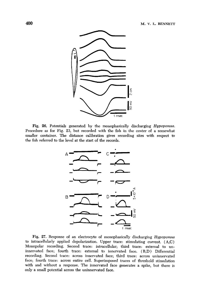
Fig. 26. Potentials generated by the monophasically discharging Hypopomus.
Procedure as for Fig. 23, but recorded with the fish in the center of a somewhat
smaller container. The distance calibration gives recording sites with respect to
the fish referred to the level at the start of the records.
a
I msec
Fig. 27. Response of an electrocyte of monophasically discharging Hypopomus
to intracellularly applied depolarization. Upper trace: stimulating current. ( A,C )
Monopolar recording. Second trace: intracellular; third trace: external to uninnervated face; fourth trace: external to innervated face. (B,D) Differential
recording. Second trace: across innervated face; third trace: across uninnervated
face; fourth trace: across entire cell. Superimposed traces of threshold stimulation
with and without a response. The innervated face generates a spike, but there is
only a small potential across the uninnervated face.
M. V. L. BENNETT
Fig. 26. Potentials generated by the monophasically discharging Hypopomus.
Procedure as for Fig. 23, but recorded with the fish in the center of a somewhat
smaller container. The distance calibration gives recording sites with respect to
the fish referred to the level at the start of the records.
a
I msec
Fig. 27. Response of an electrocyte of monophasically discharging Hypopomus
to intracellularly applied depolarization. Upper trace: stimulating current. ( A,C )
Monopolar recording. Second trace: intracellular; third trace: external to uninnervated face; fourth trace: external to innervated face. (B,D) Differential
recording. Second trace: across innervated face; third trace: across uninnervated
face; fourth trace: across entire cell. Superimposed traces of threshold stimulation
with and without a response. The innervated face generates a spike, but there is
only a small potential across the uninnervated face.