

10. ELECTRIC ORGANS
401
A=.B - n -
2 msec
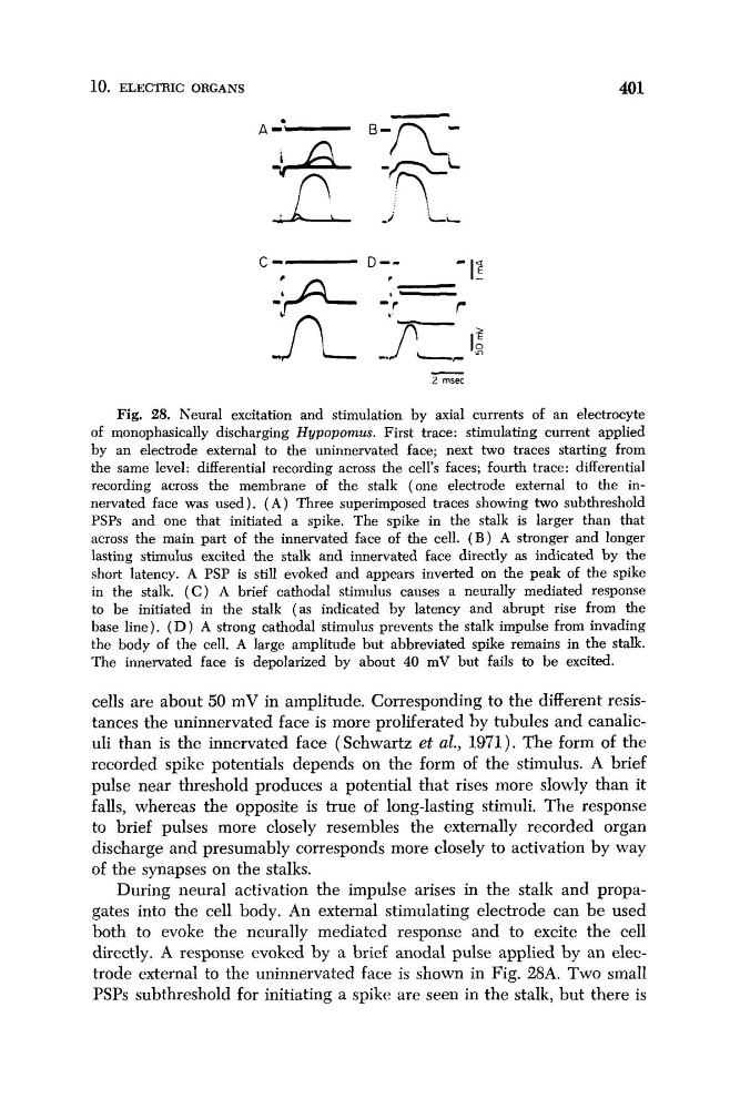
Fig. 28. Neural excitation and stimulation by axial currents of an electrocyte
of monophasically discharging Hypopomus. First trace: stimulating current applied
by an electrode external to the uninnervated face; next two traces starting from
the same level: differential recording across the cell's faces; fourth trace: differential
recording across the membrane of the stalk (one electrode external to the innervated face was used). ( A ) Three superimposed traces showing two subthreshold
PSPs and one that initiated a spike. The spike in the stalk is larger than that
across the main part of the innervated face of the cell. ( B ) A stronger and longer
lasting stimulus excited the stalk and innervated face directly as indicated by the
short latency. A PSP is still evoked and appears inverted on the peak of the spike
in the stalk. ( C ) A brief cathodal stimulus causes a neurally mediated response
to be initiated in the stalk (as indicated by latency and abrupt rise from the
base line). ( D ) A strong cathodal stimulus prevents the stalk impulse from invading
the body of the cell. A large amplitude but abbreviated spike remains in the stalk.
The innervated face is depolarized by about 40 mV but fails to be excited.
cells are about 50 mV in amplitude. Corresponding to the different resistances the uninnervated face is more proliferated by tubules and canaliculi than is the innervated face (Schwartz et al., 1971). The form of the
recorded spike potentials depends on the form of the stimulus. A brief
pulse near threshold produces a potential that rises more slowly than it
falls, whereas the opposite is true of long-lasting stimuli. The response
to brief pulses more closely resembles the externally recorded organ
discharge and presumably corresponds more closely to activation by way
of the synapses on the stalks.
During neural activation the impulse arises in the stalk and propagates into the cell body. An external stimulating electrode can be used
both to evoke the neurally mediated response and to excite the cell
directly. A response evoked by a brief anodal pulse applied by an electrode external to the uninnervated face is shown in Fig. 28A. Two small
PSPs subthreshold for initiating a spike are seen in the stalk, but there is
401
A=.B - n -
2 msec
Fig. 28. Neural excitation and stimulation by axial currents of an electrocyte
of monophasically discharging Hypopomus. First trace: stimulating current applied
by an electrode external to the uninnervated face; next two traces starting from
the same level: differential recording across the cell's faces; fourth trace: differential
recording across the membrane of the stalk (one electrode external to the innervated face was used). ( A ) Three superimposed traces showing two subthreshold
PSPs and one that initiated a spike. The spike in the stalk is larger than that
across the main part of the innervated face of the cell. ( B ) A stronger and longer
lasting stimulus excited the stalk and innervated face directly as indicated by the
short latency. A PSP is still evoked and appears inverted on the peak of the spike
in the stalk. ( C ) A brief cathodal stimulus causes a neurally mediated response
to be initiated in the stalk (as indicated by latency and abrupt rise from the
base line). ( D ) A strong cathodal stimulus prevents the stalk impulse from invading
the body of the cell. A large amplitude but abbreviated spike remains in the stalk.
The innervated face is depolarized by about 40 mV but fails to be excited.
cells are about 50 mV in amplitude. Corresponding to the different resistances the uninnervated face is more proliferated by tubules and canaliculi than is the innervated face (Schwartz et al., 1971). The form of the
recorded spike potentials depends on the form of the stimulus. A brief
pulse near threshold produces a potential that rises more slowly than it
falls, whereas the opposite is true of long-lasting stimuli. The response
to brief pulses more closely resembles the externally recorded organ
discharge and presumably corresponds more closely to activation by way
of the synapses on the stalks.
During neural activation the impulse arises in the stalk and propagates into the cell body. An external stimulating electrode can be used
both to evoke the neurally mediated response and to excite the cell
directly. A response evoked by a brief anodal pulse applied by an electrode external to the uninnervated face is shown in Fig. 28A. Two small
PSPs subthreshold for initiating a spike are seen in the stalk, but there is