

10. ELECTRIC ORGANS
397
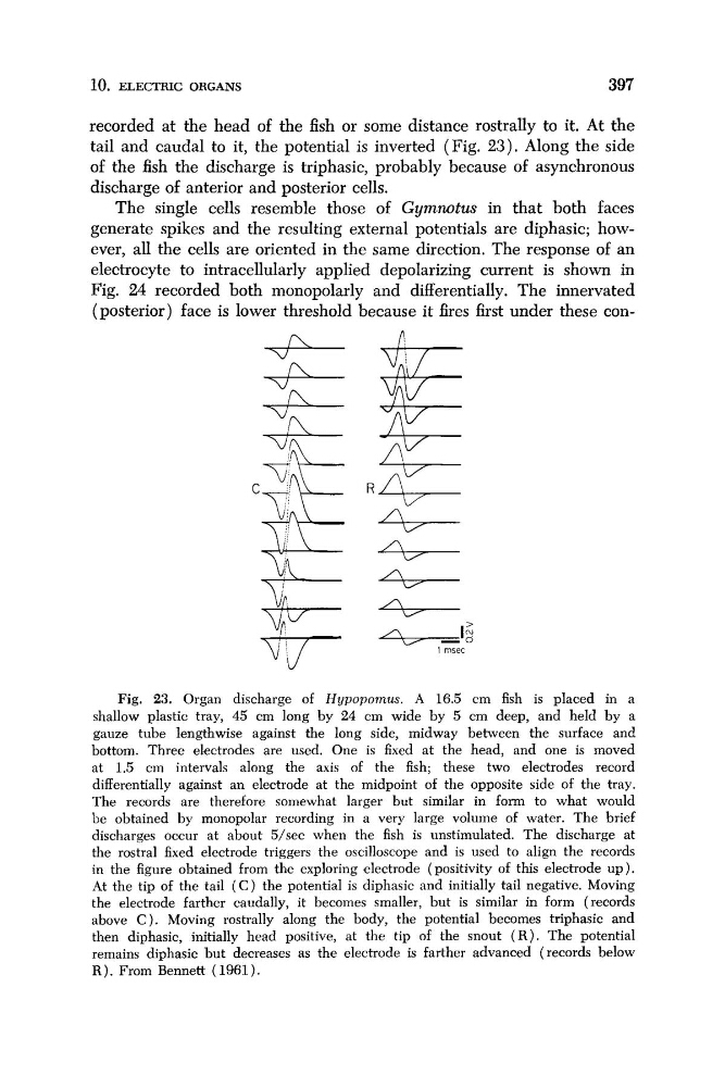
recorded at the head of the fish or some distance rostrally to it. At the
tail and caudal to it, the potential is inverted (Fig. 2 3 ) . Along the side
of the fish the discharge is triphasic, probably because of asynchronous
discharge of anterior and posterior cells.
The single cells resemble those of Gymnotus in that both faces
generate spikes and the resulting external potentials are diphasic; however, all the cells are oriented in the same direction. The response of an
electrocyte to intracellularly applied depolarizing current is shown in
Fig. 24 recorded both monopolarly and differentially. The innervated
+I rnsec
Fig. 23. Organ discharge of Hypopornus. A 16.5 cm fish is placed in a
shallow plastic tray, 45 cm long by 24 cm wide by 5 cm deep, and held by a
gauze tube lengthwise against the long side, midway between the surface and
bottom. Three electrodes are used. One is fixed at the head, and one is moved
at 1.5 cm intervals along the axis of the fish; these two electrodes record
differentially against an electrode at the midpoint of the opposite side of the tray.
The records are therefore somewhat larger but similar in form to what would
be obtained by monopolar recording in a very large volume of water. The brief
discharges occur at about 5/sec when the fish is unstimulated. The discharge at
the rostra1 fixed electrode triggers the oscilloscope and is used to align the records
in the figure obtained from the exploring electrode (positivity of this electrode up).
At the tip of the tail ( C ) the potential is diphasic and initially tail negative. Moving
the electrode farther caudally, it becomes smaller, but is similar in form (records
above C ) . Moving rostrally along the body, the potential becomes triphasic and
then diphasic, initially head positive, at the tip of the snout ( R ) . The potential
remains diphasic but decreases as the electrode is farther advanced (records below
R ) . From Bennett (1961).
397
recorded at the head of the fish or some distance rostrally to it. At the
tail and caudal to it, the potential is inverted (Fig. 2 3 ) . Along the side
of the fish the discharge is triphasic, probably because of asynchronous
discharge of anterior and posterior cells.
The single cells resemble those of Gymnotus in that both faces
generate spikes and the resulting external potentials are diphasic; however, all the cells are oriented in the same direction. The response of an
electrocyte to intracellularly applied depolarizing current is shown in
Fig. 24 recorded both monopolarly and differentially. The innervated
+I rnsec
Fig. 23. Organ discharge of Hypopornus. A 16.5 cm fish is placed in a
shallow plastic tray, 45 cm long by 24 cm wide by 5 cm deep, and held by a
gauze tube lengthwise against the long side, midway between the surface and
bottom. Three electrodes are used. One is fixed at the head, and one is moved
at 1.5 cm intervals along the axis of the fish; these two electrodes record
differentially against an electrode at the midpoint of the opposite side of the tray.
The records are therefore somewhat larger but similar in form to what would
be obtained by monopolar recording in a very large volume of water. The brief
discharges occur at about 5/sec when the fish is unstimulated. The discharge at
the rostra1 fixed electrode triggers the oscilloscope and is used to align the records
in the figure obtained from the exploring electrode (positivity of this electrode up).
At the tip of the tail ( C ) the potential is diphasic and initially tail negative. Moving
the electrode farther caudally, it becomes smaller, but is similar in form (records
above C ) . Moving rostrally along the body, the potential becomes triphasic and
then diphasic, initially head positive, at the tip of the snout ( R ) . The potential
remains diphasic but decreases as the electrode is farther advanced (records below
R ) . From Bennett (1961).