

396
M. V. L. BENNElT
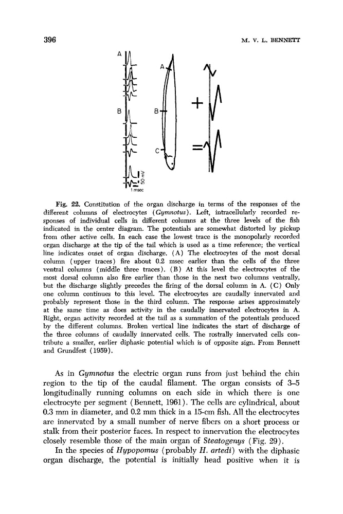
Fig. 22. Constitution of the organ discharge in terms of the responses of the
different columns of electrocytes ( Gymnotus). Left, intracellularly recorded responses of individual cells in different columns at the three levels of the fish
indicated in the center diagram. The potentials are somewhat distorted by pickup
from other active cells. In each case the lowest trace is the monopolarly recorded
organ discharge at the tip of the tail which is used as a time reference; the vertical
line indicates onset of organ discharge. ( A ) The electrocytes of the most dorsal
column (upper traces) fire about 0.2 msec earlier than the cells of the three
ventral columns (middle three traces). ( B ) At this level the electrocytes of the
most dorsal column also fire earlier than those in the next two columns ventrally,
but the discharge slightly precedes the firing of the dorsal column in A. ( C ) Only
one column continues to this level. The electrocytes are caudally innervated and
probably represent those in the third column. The response arises approximately
at the same time as does activity in the caudally innervated electrocytes in A.
Right, organ activity recorded at the tail as a summation of the potentials produced
by the different columns. Broken vertical line indicates the start of discharge of
the three columns of caudally innervated cells. The rostrally innervated cells contribute a smaller, earlier diphasic potential which is of opposite sign. From Bennett
and Grundfest (1959).
As in Gymnotus the electric organ runs from just behind the chin
region to the tip of the caudal filament. The organ consists of 3-5
longitudinally running columns on each side in which there is one
electrocyte per segment ( Bennett, 1961). The cells are cylindrical, about
0.3 mm in diameter, and 0.2 mm thick in a 15-cm fish. All the electrocytes
are innervated by a small number of nerve fibers on a short process or
stalk from their posterior faces. In respect to innervation the electrocytes
closely resemble those of the main organ of Steatogenys (Fig. 29).
In the species of Hypopomus (probably H . urtedi) with the diphasic
organ discharge, the potential is initially head positive when it is
M. V. L. BENNElT
Fig. 22. Constitution of the organ discharge in terms of the responses of the
different columns of electrocytes ( Gymnotus). Left, intracellularly recorded responses of individual cells in different columns at the three levels of the fish
indicated in the center diagram. The potentials are somewhat distorted by pickup
from other active cells. In each case the lowest trace is the monopolarly recorded
organ discharge at the tip of the tail which is used as a time reference; the vertical
line indicates onset of organ discharge. ( A ) The electrocytes of the most dorsal
column (upper traces) fire about 0.2 msec earlier than the cells of the three
ventral columns (middle three traces). ( B ) At this level the electrocytes of the
most dorsal column also fire earlier than those in the next two columns ventrally,
but the discharge slightly precedes the firing of the dorsal column in A. ( C ) Only
one column continues to this level. The electrocytes are caudally innervated and
probably represent those in the third column. The response arises approximately
at the same time as does activity in the caudally innervated electrocytes in A.
Right, organ activity recorded at the tail as a summation of the potentials produced
by the different columns. Broken vertical line indicates the start of discharge of
the three columns of caudally innervated cells. The rostrally innervated cells contribute a smaller, earlier diphasic potential which is of opposite sign. From Bennett
and Grundfest (1959).
As in Gymnotus the electric organ runs from just behind the chin
region to the tip of the caudal filament. The organ consists of 3-5
longitudinally running columns on each side in which there is one
electrocyte per segment ( Bennett, 1961). The cells are cylindrical, about
0.3 mm in diameter, and 0.2 mm thick in a 15-cm fish. All the electrocytes
are innervated by a small number of nerve fibers on a short process or
stalk from their posterior faces. In respect to innervation the electrocytes
closely resemble those of the main organ of Steatogenys (Fig. 29).
In the species of Hypopomus (probably H . urtedi) with the diphasic
organ discharge, the potential is initially head positive when it is