

9. THE MAUTHNER CELL
327
Hyper polar izot ion
L
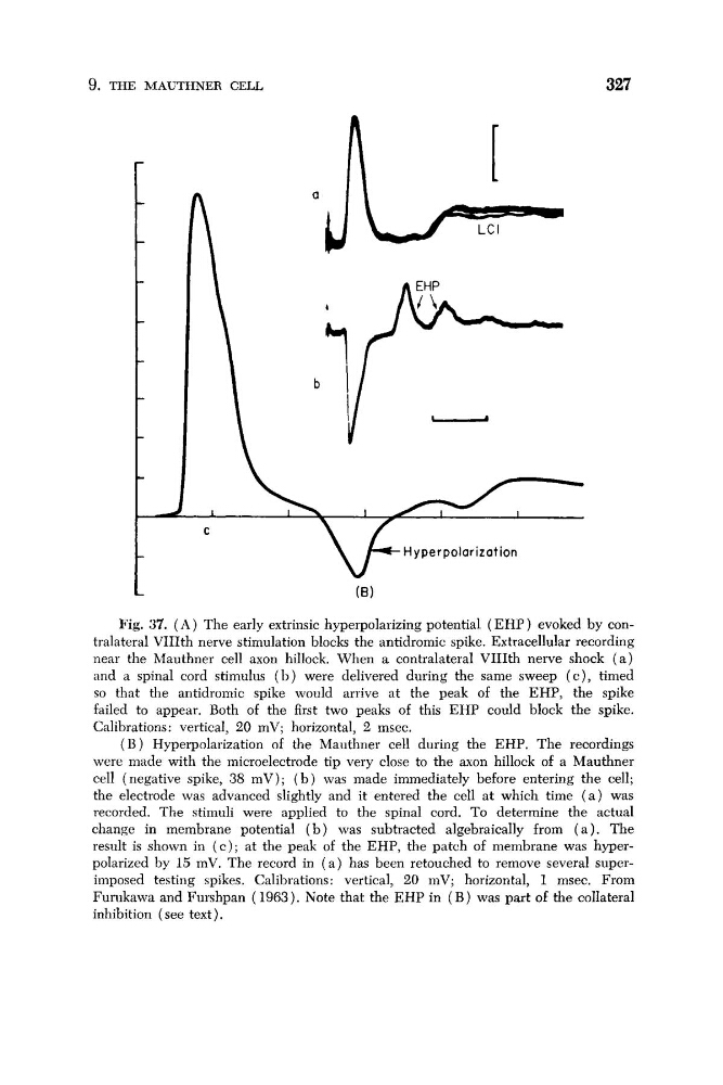
Fig. 37. ( A ) The early extrinsic hyperpolarizing potential ( E H P ) evoked by contralateral VIIIth nerve stimulation blocks the antidromic spike. Extracellular recording
near the Mauthner cell axon hillock. When a contralateral VIIIth nerve shock ( a )
and a spinal cord stimulus (11) were delivered during the same sweep ( c ) , timed
so that the antidroniic spike would arrive at the peak of the EHP, the spike
failed to appear. Both of the first two peaks of this EHP could block the spike.
Calibrations: vertical, 20 mV; horizontal, 2 msec.
( B ) Hyperpolarization of the Mauthner cell during the EHP. The recordings
were made with the microelectrode tip very close to the axon hillock of a Mauthner
cell (negative spike, 38 mV); ( b ) was made immediately before entering the cell;
the electrode was advanced slightly and it entered the cell at which time ( a ) was
recorded. The stimuli were applied to the spinal cord. To determine the actual
change in membrane potential ( b ) was subtracted algebraically from ( a ) . The
result is shown in ( c ) ; at the peak of the EHP, the patch of membrane was hyperpolarized by 15 mV. The record in ( a ) has been retouched to remove several superimposed testing spikes. Calibrations: vertical, 20 mV; horizontal, 1 msec. From
Furukawa and Furshpan ( 1963). Note that the EHP in ( B ) was part of the collateral
inhibition (see text).
327
Hyper polar izot ion
L
Fig. 37. ( A ) The early extrinsic hyperpolarizing potential ( E H P ) evoked by contralateral VIIIth nerve stimulation blocks the antidromic spike. Extracellular recording
near the Mauthner cell axon hillock. When a contralateral VIIIth nerve shock ( a )
and a spinal cord stimulus (11) were delivered during the same sweep ( c ) , timed
so that the antidroniic spike would arrive at the peak of the EHP, the spike
failed to appear. Both of the first two peaks of this EHP could block the spike.
Calibrations: vertical, 20 mV; horizontal, 2 msec.
( B ) Hyperpolarization of the Mauthner cell during the EHP. The recordings
were made with the microelectrode tip very close to the axon hillock of a Mauthner
cell (negative spike, 38 mV); ( b ) was made immediately before entering the cell;
the electrode was advanced slightly and it entered the cell at which time ( a ) was
recorded. The stimuli were applied to the spinal cord. To determine the actual
change in membrane potential ( b ) was subtracted algebraically from ( a ) . The
result is shown in ( c ) ; at the peak of the EHP, the patch of membrane was hyperpolarized by 15 mV. The record in ( a ) has been retouched to remove several superimposed testing spikes. Calibrations: vertical, 20 mV; horizontal, 1 msec. From
Furukawa and Furshpan ( 1963). Note that the EHP in ( B ) was part of the collateral
inhibition (see text).