

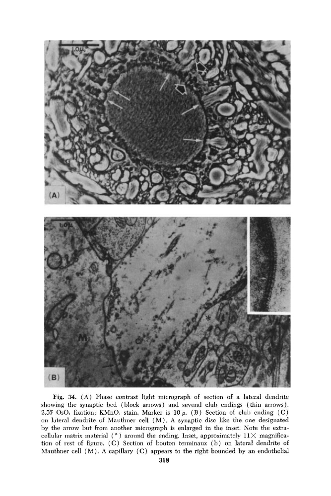
Fig. 34. ( A ) Phase contrast light micrograph of section of a lateral dendrite
showing the synaptic bed (block arrows) and several club endings (thin arrows).
2.5% OsO, fixation; KMnOI stain. Marker is 10 p. ( B ) Section of club ending ( C )
on lateral dendrite of Mauthner cell ( M ) . A synaptic disc like the one designated
by the arrow but from another micrograph is enlarged in the inset. Note the extracellular inatrix material ( * ) around the ending. Inset, approximately 11 X magnification of rest of figure. ( C ) Section of bouton terniinaux ( b ) on lateral dendrite of
Mauthner cell ( M ) . A capillary ( C ) appears to the right bounded by an endothelial
318
showing the synaptic bed (block arrows) and several club endings (thin arrows).
2.5% OsO, fixation; KMnOI stain. Marker is 10 p. ( B ) Section of club ending ( C )
on lateral dendrite of Mauthner cell ( M ) . A synaptic disc like the one designated
by the arrow but from another micrograph is enlarged in the inset. Note the extracellular inatrix material ( * ) around the ending. Inset, approximately 11 X magnification of rest of figure. ( C ) Section of bouton terniinaux ( b ) on lateral dendrite of
Mauthner cell ( M ) . A capillary ( C ) appears to the right bounded by an endothelial
318