

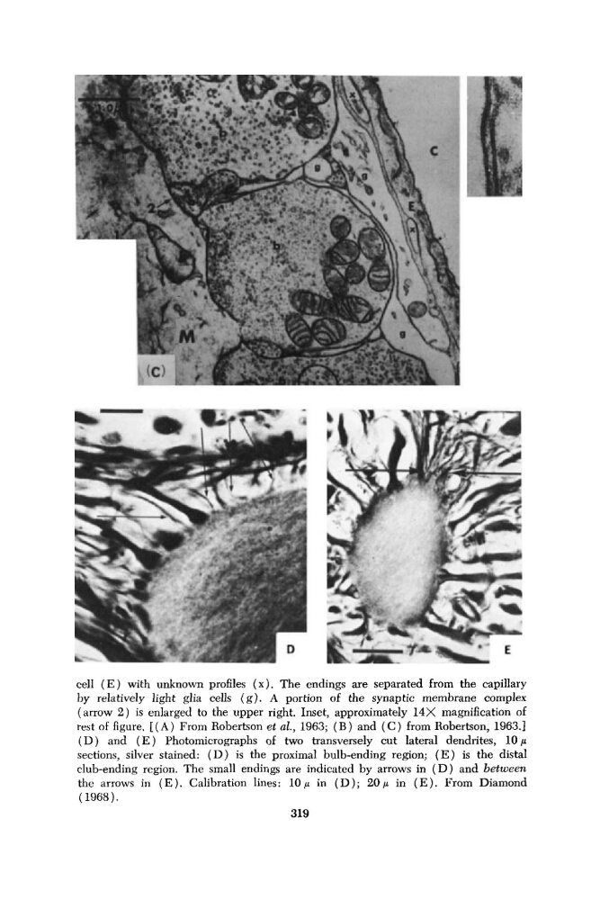
cell ( E ) with unknown profiles (x). The endings are separated from the capillary
by relatively light glia cells (g). A portion of the synaptic membrane complex
(arrow 2 ) is enlarged to the upper right. Inset, approximately 14X magnification of
rest of figure. [ ( A ) From Robertson et al., 1963; ( B ) and ( C ) from Robertson, 1963.1
( D ) and ( E ) Photomicrographs of two transversely cut lateral dendrites, 10p
sections, silver stained: ( D ) is the proximal bulb-ending region; ( E ) is the distal
club-ending region. The small endings are indicated by arrows in ( D ) and between
the arrows in ( E ). Calibration lines: 10 p in ( D ) ; 20 p in ( E ). From Diamond
(1968).
319
by relatively light glia cells (g). A portion of the synaptic membrane complex
(arrow 2 ) is enlarged to the upper right. Inset, approximately 14X magnification of
rest of figure. [ ( A ) From Robertson et al., 1963; ( B ) and ( C ) from Robertson, 1963.1
( D ) and ( E ) Photomicrographs of two transversely cut lateral dendrites, 10p
sections, silver stained: ( D ) is the proximal bulb-ending region; ( E ) is the distal
club-ending region. The small endings are indicated by arrows in ( D ) and between
the arrows in ( E ). Calibration lines: 10 p in ( D ) ; 20 p in ( E ). From Diamond
(1968).
319