

9. THE MAUTHNER CELL
317
-
O.5msec
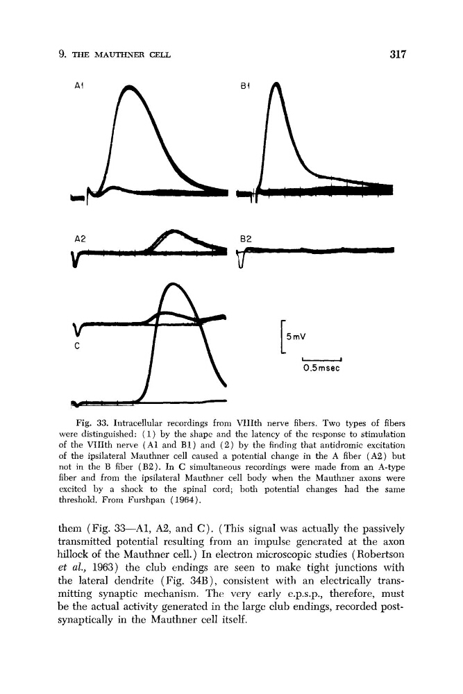
Fig. 33. Intracellular recordings from VIIIth nerve fibers. Two types of fibers
were distinguished: ( 1) by the shape and the latency of the response to stimulation
of the VIIIth nerve ( A 1 and B1) and ( 2 ) by the finding that antidromic excitation
of the ipsilateral Mauthner cell caused a potential change in the A fiber ( A 2 ) but
not in the B fiber ( B 2 ) . In C simultaneous recordings were made from an A-type
fiber and from the ipsilateral Mauthner cell body when the Mauthner axons were
excited by a shock to the spinal cord; both potential changes had the same
threshold. From Furshpan (1964).
them (Fig. 33-A1, A2, and C ) . (This signal was actually the passively
transmitted potential resulting from an impulse generated at the axon
hillock of the Mauthner cell.) In electron microscopic studies (Robertson
et al., 1963) the club endings are seen to make tight junctions with
the lateral dendrite (Fig. 34B), consistent with an electrically transmitting synaptic mechanism. The very early e.p.s.p., therefore, must
be the actual activity generated in the large club endings, recorded postsynaptically in the Mauthner cell itself.
317
-
O.5msec
Fig. 33. Intracellular recordings from VIIIth nerve fibers. Two types of fibers
were distinguished: ( 1) by the shape and the latency of the response to stimulation
of the VIIIth nerve ( A 1 and B1) and ( 2 ) by the finding that antidromic excitation
of the ipsilateral Mauthner cell caused a potential change in the A fiber ( A 2 ) but
not in the B fiber ( B 2 ) . In C simultaneous recordings were made from an A-type
fiber and from the ipsilateral Mauthner cell body when the Mauthner axons were
excited by a shock to the spinal cord; both potential changes had the same
threshold. From Furshpan (1964).
them (Fig. 33-A1, A2, and C ) . (This signal was actually the passively
transmitted potential resulting from an impulse generated at the axon
hillock of the Mauthner cell.) In electron microscopic studies (Robertson
et al., 1963) the club endings are seen to make tight junctions with
the lateral dendrite (Fig. 34B), consistent with an electrically transmitting synaptic mechanism. The very early e.p.s.p., therefore, must
be the actual activity generated in the large club endings, recorded postsynaptically in the Mauthner cell itself.