

J. DIAMOND
304
M a
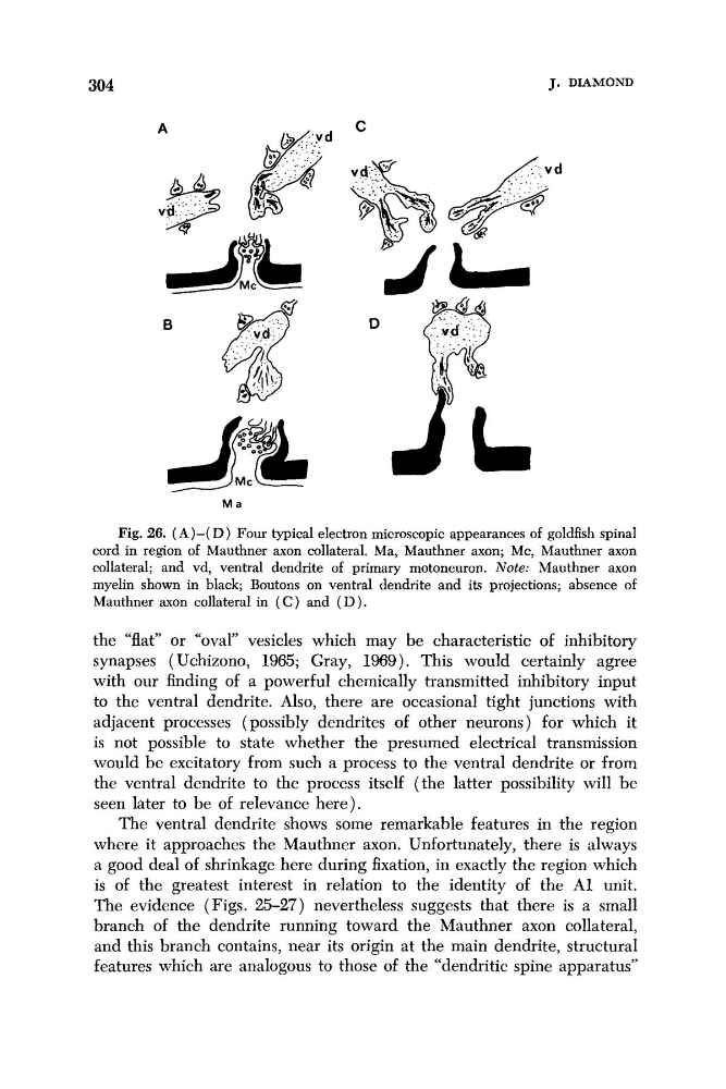
Fig. 26. (A)-( D ) Four typical electron microscopic appearances of goldfish spinal
cord in region of Mauthner axon collateral. Ma, Mauthner axon; Mc, Mauthner axon
collateral; and vd, ventral dendrite of primary motoneuron. Note: Mauthner axon
myelin shown in black; Boutons on ventral dendrite and its projections; absence of
Mauthner axon collateral in ( C ) and ( D ) .
the “flat” or “oval” vesicles which may be characteristic of inhibitory
synapses ( Uchizono, 1965; Gray, 1969). This would certainly agree
with our finding of a powerful chemically transmitted inhibitory input
to the ventral dendrite. Also, there are occasional tight junctions with
adjacent processes (possibly dendrites of other neurons) for which it
is not possible to state whether the presumed electrical transmission
would be excitatory from such a process to the ventral dendrite or from
the ventral dendrite to the process itself (the latter possibility will be
seen later to be of relevance here).
The ventral dendrite shows some remarkable features in the region
where it approaches the Mauthner axon. Unfortunately, there is always
a good deal of shrinkage here during fixation, in exactly the region which
is of the greatest interest in relation to the identity of the A1 unit.
The evidence (Figs. 25-27) nevertheless suggests that there is a small
branch of the dendrite running toward the Mauthner axon collateral,
and this branch contains, near its origin at the main dendrite, structural
features which are analogous to those of the “dendritic spine apparatus”
304
M a
Fig. 26. (A)-( D ) Four typical electron microscopic appearances of goldfish spinal
cord in region of Mauthner axon collateral. Ma, Mauthner axon; Mc, Mauthner axon
collateral; and vd, ventral dendrite of primary motoneuron. Note: Mauthner axon
myelin shown in black; Boutons on ventral dendrite and its projections; absence of
Mauthner axon collateral in ( C ) and ( D ) .
the “flat” or “oval” vesicles which may be characteristic of inhibitory
synapses ( Uchizono, 1965; Gray, 1969). This would certainly agree
with our finding of a powerful chemically transmitted inhibitory input
to the ventral dendrite. Also, there are occasional tight junctions with
adjacent processes (possibly dendrites of other neurons) for which it
is not possible to state whether the presumed electrical transmission
would be excitatory from such a process to the ventral dendrite or from
the ventral dendrite to the process itself (the latter possibility will be
seen later to be of relevance here).
The ventral dendrite shows some remarkable features in the region
where it approaches the Mauthner axon. Unfortunately, there is always
a good deal of shrinkage here during fixation, in exactly the region which
is of the greatest interest in relation to the identity of the A1 unit.
The evidence (Figs. 25-27) nevertheless suggests that there is a small
branch of the dendrite running toward the Mauthner axon collateral,
and this branch contains, near its origin at the main dendrite, structural
features which are analogous to those of the “dendritic spine apparatus”