

9. THE MAUTHNER CELL
303
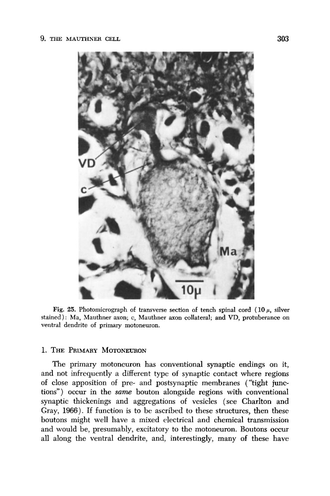
Fig. 25. Photomicrograph of transverse section of tench spinal cord (10 p, silver
stained): Ma, Mauthner axon; c, Mauthner axon collateral; and VD, protuberance on
ventral dendrite of primary motoneuron.
1. THE PRIMARY MOTONEURON
The primary motoneuron has conventional synaptic endings on it,
and not infrequently a different type of synaptic contact where regions
of close apposition of pre- and postsynaptic membranes (“tight junctions”) occur in the same bouton alongside regions with conventional
synaptic thickenings and aggregations of vesicles (see Charlton and
Gray, 1966). If function is to be ascribed to these structures, then these
boutons might well have a mixed electrical and chemical transmission
and would be, presumably, excitatory to the motoneuron. Boutons occur
all along the ventral dendrite, and, interestingly, many of these have
303
Fig. 25. Photomicrograph of transverse section of tench spinal cord (10 p, silver
stained): Ma, Mauthner axon; c, Mauthner axon collateral; and VD, protuberance on
ventral dendrite of primary motoneuron.
1. THE PRIMARY MOTONEURON
The primary motoneuron has conventional synaptic endings on it,
and not infrequently a different type of synaptic contact where regions
of close apposition of pre- and postsynaptic membranes (“tight junctions”) occur in the same bouton alongside regions with conventional
synaptic thickenings and aggregations of vesicles (see Charlton and
Gray, 1966). If function is to be ascribed to these structures, then these
boutons might well have a mixed electrical and chemical transmission
and would be, presumably, excitatory to the motoneuron. Boutons occur
all along the ventral dendrite, and, interestingly, many of these have