

9. THE MAUTHNER CELL
305
I
Mauthner axon
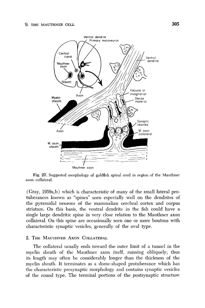
Fig. 27. Suggested morphology of goldfish spinal cord in region of the Mauthner
axon collateral.
(Gray, 1959a,b) which is characteristic of many of the small lateral protuberances known as “spines” seen especially well on the dendrites of
the pyramidal neurons of the mammalian cerebral cortex and corpus
striatuni. On this basis, the ventral dendrite in the fish could have a
single large dendritic spine in very close relation to the Mauthner axon
collateral. On this spine are occasionally seen one or more boutons with
characteristic synaptic vesicles, generally of the oval type.
2. THE MAUTHNER AXON COLLATERAL
The collateral usually ends toward the outer limit of a tunnel in the
myelin sheath of the Mauthner axon itself, running obliquely; thus
its length may often be considerably longer than the thickness of the
myelin sheath. It terminates as a dome-shaped protuberance which has
the characteristic presynaptic morphology and contains synaptic vesicles
of the round type. The terminal portions of the postsynaptic structure
305
I
Mauthner axon
Fig. 27. Suggested morphology of goldfish spinal cord in region of the Mauthner
axon collateral.
(Gray, 1959a,b) which is characteristic of many of the small lateral protuberances known as “spines” seen especially well on the dendrites of
the pyramidal neurons of the mammalian cerebral cortex and corpus
striatuni. On this basis, the ventral dendrite in the fish could have a
single large dendritic spine in very close relation to the Mauthner axon
collateral. On this spine are occasionally seen one or more boutons with
characteristic synaptic vesicles, generally of the oval type.
2. THE MAUTHNER AXON COLLATERAL
The collateral usually ends toward the outer limit of a tunnel in the
myelin sheath of the Mauthner axon itself, running obliquely; thus
its length may often be considerably longer than the thickness of the
myelin sheath. It terminates as a dome-shaped protuberance which has
the characteristic presynaptic morphology and contains synaptic vesicles
of the round type. The terminal portions of the postsynaptic structure