

9. THE MAUTHNER CELL
295
I'
b
-.
C
d
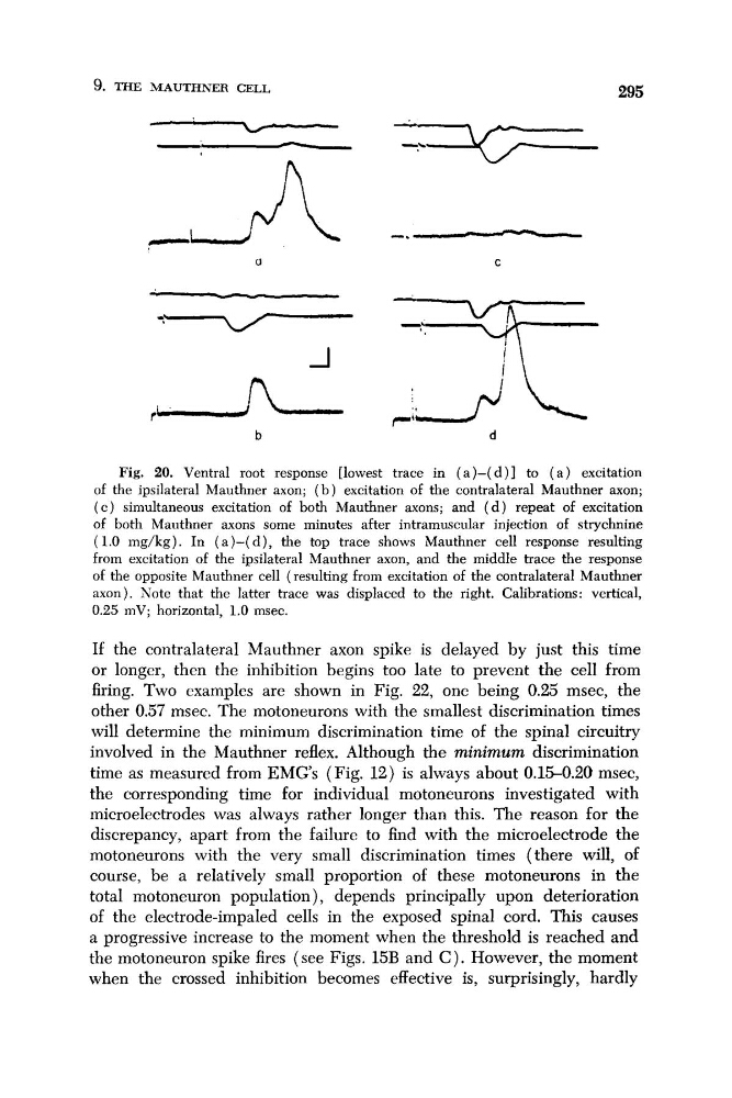
Fig. 20. Ventral root response [lowest trace in ( a ) - ( d ) ] to ( a ) excitation
of the ipsilateral Mauthner axon; ( b ) excitation of the contralateral Mauthner axon;
( c ) simultaneous excitation of both Mauthner axons; and ( d ) repeat of excitation
of both Mauthner axons some minutes after intramuscular injection of strychnine
(1.0 mg/kg). In (a)-( d ) , the top trace shows Mauthner cell response resulting
from excitation of the ipsilateral Mauthner axon, and the middle trace the response
of the opposite Mauthner cell (resulting from excitation of the contralateral Mauthner
axon). Note that the latter trace was displaced to the right. Calibrations: vertical,
0.25 mV; horizontal, 1.0 msec.
If the contralateral Mauthner axon spike is delayed by just this time
or longer, then the inhibition begins too late to prevent the cell from
firing. Two examples are shown in Fig. 22, one being 0.25 msec, the
other 0.57 msec. The motoneurons with the smallest discrimination times
will determine the minimum discrimination time of the spinal circuitry
involved in the Mauthner reflex. Although the minimum discrimination
time as measured from EMGs (Fig. 12) is always about 0.15-0.20 msec,
the corresponding time for individual motoneurons investigated with
microelectrodes was always rather longer than this. The reason for the
discrepancy, apart from the failure to find with the microelectrode the
motoneurons with the very small discrimination times (there will, of
course, be a relatively small proportion of these motoneurons in the
total motoneuron population), depends principally upon deterioration
of the electrode-impaled cells in the exposed spinal cord. This causes
a progressive increase to the moment when the threshold is reached and
the motoneuron spike fires (see Figs. 15B and C). However, the moment
when the crossed inhibition becomes effective is, surprisingly, hardly
295
I'
b
-.
C
d
Fig. 20. Ventral root response [lowest trace in ( a ) - ( d ) ] to ( a ) excitation
of the ipsilateral Mauthner axon; ( b ) excitation of the contralateral Mauthner axon;
( c ) simultaneous excitation of both Mauthner axons; and ( d ) repeat of excitation
of both Mauthner axons some minutes after intramuscular injection of strychnine
(1.0 mg/kg). In (a)-( d ) , the top trace shows Mauthner cell response resulting
from excitation of the ipsilateral Mauthner axon, and the middle trace the response
of the opposite Mauthner cell (resulting from excitation of the contralateral Mauthner
axon). Note that the latter trace was displaced to the right. Calibrations: vertical,
0.25 mV; horizontal, 1.0 msec.
If the contralateral Mauthner axon spike is delayed by just this time
or longer, then the inhibition begins too late to prevent the cell from
firing. Two examples are shown in Fig. 22, one being 0.25 msec, the
other 0.57 msec. The motoneurons with the smallest discrimination times
will determine the minimum discrimination time of the spinal circuitry
involved in the Mauthner reflex. Although the minimum discrimination
time as measured from EMGs (Fig. 12) is always about 0.15-0.20 msec,
the corresponding time for individual motoneurons investigated with
microelectrodes was always rather longer than this. The reason for the
discrepancy, apart from the failure to find with the microelectrode the
motoneurons with the very small discrimination times (there will, of
course, be a relatively small proportion of these motoneurons in the
total motoneuron population), depends principally upon deterioration
of the electrode-impaled cells in the exposed spinal cord. This causes
a progressive increase to the moment when the threshold is reached and
the motoneuron spike fires (see Figs. 15B and C). However, the moment
when the crossed inhibition becomes effective is, surprisingly, hardly