

290
1 23
: ! / I
J. DIAMOND
A 2 y T
(DETERIORATEDIi
(C) -
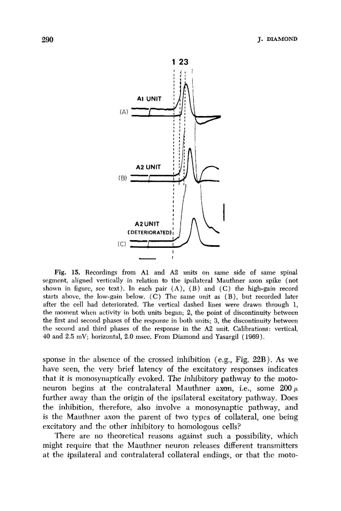
Fig. 15. Recordings from A1 and A2 units on same side of same spinal
segment, aligned vertically in relation to the ipsilateral Mauthner axon spike (not
shown in figure, see text). In each pair ( A ) , ( B ) and ( C ) the high-gain record
starts above, the low-gain below, ( C ) The same unit as ( B ) , but recorded later
after the cell had deteriorated. The vertical dashed lines were drawn through 1,
the moment when activity in both units began; 2, the point of discontinuity between
the first and second phases of the response in both units; 3, the discontinuity between
the second and third phases of the response in the A2 unit. Calibrations: vertical,
40 and 2.5 mV; horizontal, 2.0 msec. From Diamond and Yasargil (1969).
sponse in the absence of the crossed inhibition (e.g., Fig. 22B). As we
have seen, the very brief latency of the excitatory responses indicates
that it is monosynaptically evoked. The inhibitory pathway to the motoneuron begins at the contralateral Mauthner axon, i.e., some 2 0 0 p
further away than the origin of the ipsilateral excitatory pathway. Does
the inhibition, therefore, also involve a monosynaptic pathway, and
is the Mauthner axon the parent of two types of collateral, one being
excitatory and the other inhibitory to homologous cells?
There are no theoretical reasons against such a possibility, which
might require that the Mauthner neuron releases different transmitters
at the ipsilateral and contralateral collateral endings, or that the moto-
1 23
: ! / I
J. DIAMOND
A 2 y T
(DETERIORATEDIi
(C) -
Fig. 15. Recordings from A1 and A2 units on same side of same spinal
segment, aligned vertically in relation to the ipsilateral Mauthner axon spike (not
shown in figure, see text). In each pair ( A ) , ( B ) and ( C ) the high-gain record
starts above, the low-gain below, ( C ) The same unit as ( B ) , but recorded later
after the cell had deteriorated. The vertical dashed lines were drawn through 1,
the moment when activity in both units began; 2, the point of discontinuity between
the first and second phases of the response in both units; 3, the discontinuity between
the second and third phases of the response in the A2 unit. Calibrations: vertical,
40 and 2.5 mV; horizontal, 2.0 msec. From Diamond and Yasargil (1969).
sponse in the absence of the crossed inhibition (e.g., Fig. 22B). As we
have seen, the very brief latency of the excitatory responses indicates
that it is monosynaptically evoked. The inhibitory pathway to the motoneuron begins at the contralateral Mauthner axon, i.e., some 2 0 0 p
further away than the origin of the ipsilateral excitatory pathway. Does
the inhibition, therefore, also involve a monosynaptic pathway, and
is the Mauthner axon the parent of two types of collateral, one being
excitatory and the other inhibitory to homologous cells?
There are no theoretical reasons against such a possibility, which
might require that the Mauthner neuron releases different transmitters
at the ipsilateral and contralateral collateral endings, or that the moto-