

9. THE MAUTHNER CELL
289
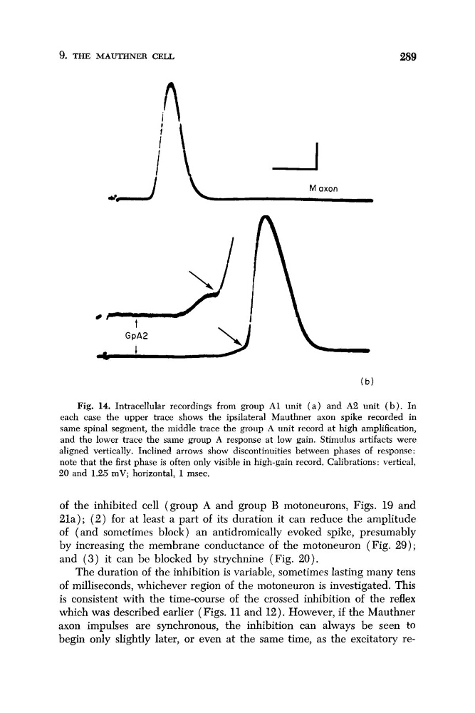
Fig. 14. Intracellular recordings from group A1 unit ( a ) and A2 unit ( b ) . In
each case the upper trace shows the ipsilateral Mauthner axon spike recorded in
same spinal segment, the middle trace the group A unit record at high amplification,
and the lower trace the same group A response at low gain. Stimulus artifacts were
aligned vertically. Inclined arrows show discontinuities between phases of re'iponse:
note that the first phase is often only visible in high-gain record. Calibrations: vertical,
20 and 1.25 mV; horizontal, 1 msec.
of the inhibited cell (group A and group B motoneurons, Figs. 19' and
21a); ( 2 ) for at least a part of its duration it can reduce the amplitude
of ( and sometimes block) an antidromically evoked spike, presumably
by increasing the membrane conductance of the motoneuron (Fig. 29) ;
and ( 3 ) it can be blocked by strychnine (Fig. 20).
The duration of the inhibition is variable, sometimes lasting many tens
of milliseconds, whichever region of the motoneuron is investigated. This
is consistent with the time-course of the crossed inhibition of the reflex
which was described earlier (Figs. 11 and 12). However, if the Mauthner
axon impulses are synchronous, the inhibition can always be seen to
begin only slightly later, or even at the same time, as the excitatory re-
289
Fig. 14. Intracellular recordings from group A1 unit ( a ) and A2 unit ( b ) . In
each case the upper trace shows the ipsilateral Mauthner axon spike recorded in
same spinal segment, the middle trace the group A unit record at high amplification,
and the lower trace the same group A response at low gain. Stimulus artifacts were
aligned vertically. Inclined arrows show discontinuities between phases of re'iponse:
note that the first phase is often only visible in high-gain record. Calibrations: vertical,
20 and 1.25 mV; horizontal, 1 msec.
of the inhibited cell (group A and group B motoneurons, Figs. 19' and
21a); ( 2 ) for at least a part of its duration it can reduce the amplitude
of ( and sometimes block) an antidromically evoked spike, presumably
by increasing the membrane conductance of the motoneuron (Fig. 29) ;
and ( 3 ) it can be blocked by strychnine (Fig. 20).
The duration of the inhibition is variable, sometimes lasting many tens
of milliseconds, whichever region of the motoneuron is investigated. This
is consistent with the time-course of the crossed inhibition of the reflex
which was described earlier (Figs. 11 and 12). However, if the Mauthner
axon impulses are synchronous, the inhibition can always be seen to
begin only slightly later, or even at the same time, as the excitatory re-