

9. THE MAUTHNER CELL
277
Mauthner axon and by the second pair of the other. When orthodromic excitation was required, similar pairs of electrodes were inseited through holes
in the skull; thus, the electrode tips were close to the respective VIIIth
nerves in the region where the nerve bundles fan out near the vestibular
apparatus.
For recording, conventional microelectrodes were positioned deep in the
exposed medulla by micromanipulators; thus, their tips were near enough
to the respective Mauthner cells on either side to record the characteristic
axon hillock spikes when they were excited. Similar microelectrodes were
used to record the activity of cells in the spinal cord. Muscle activity was
also investigated; concentric needle electrodes recorded the electromyogram
(EMG) of muscles on each side of the trunk or tail, in both retro-orbital
regions, and of opercular muscles of either side. Ventral root activity was
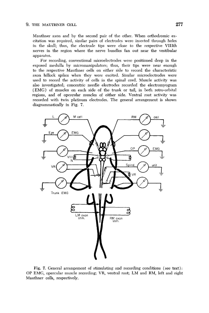
recorded with twin platinum electrodes. The general arrangement is shown
diagrammatically in Fig. 7.
Fig. 7. General arrangement of stimulating and recording conditions (see text) :
OP EMG, opercular muscle recording; VR, ventral root; LM and RM, left and right
Mauthner cells, respectively.
277
Mauthner axon and by the second pair of the other. When orthodromic excitation was required, similar pairs of electrodes were inseited through holes
in the skull; thus, the electrode tips were close to the respective VIIIth
nerves in the region where the nerve bundles fan out near the vestibular
apparatus.
For recording, conventional microelectrodes were positioned deep in the
exposed medulla by micromanipulators; thus, their tips were near enough
to the respective Mauthner cells on either side to record the characteristic
axon hillock spikes when they were excited. Similar microelectrodes were
used to record the activity of cells in the spinal cord. Muscle activity was
also investigated; concentric needle electrodes recorded the electromyogram
(EMG) of muscles on each side of the trunk or tail, in both retro-orbital
regions, and of opercular muscles of either side. Ventral root activity was
recorded with twin platinum electrodes. The general arrangement is shown
diagrammatically in Fig. 7.
Fig. 7. General arrangement of stimulating and recording conditions (see text) :
OP EMG, opercular muscle recording; VR, ventral root; LM and RM, left and right
Mauthner cells, respectively.