

276
J. DIAMOND
P
P
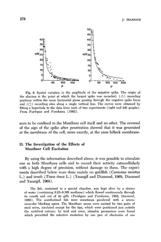
Fig. 6. Spatial variation in the amplitude of the negative spike. The origin of
the abscissa is the point at which the largest spike was recorded: ( A ) recording
positions within the same horizontal plane passing through the negative spike focus
and ( 0) recording sites along a single vertical line. The curves were obtained by
fitting a hyperbola to the data from each of two experiments (right and left graphs).
From Furshpan and Furukawa (1962).
seen to be confined to the Mauthner cell itself and no other. The reversal
of the sign of the spike after penetration showed that it was generated
at the membrane of the cell, more exactly, at the axon hillock membrane.
D. The Investigation of the Effects of
Mauthner Cell Excitation
By using the information described above, it was possible to stimulate
one or both Mauthner cells and to record their activity extracellularly
with a high degree of precision, without damage to them. The experiments described below were done mainly on goldfish (Carassius auratus
L.) and tench (Tincu tinca L.) (Yasargil and Diamond, 1968; Diamond
and Yasargil, 1969).
The fish, contained in a special chamber, was kept alive by a stream
of water (containing 0.25-0.30% urethane) which flowed continuously through
its mouth and out of its gills (Furshpan and Furukawa, 1962; Diamond,
1968). The anesthetized fish were sometimes paralyzed with a neuromuscular blocking agent. The Mauthner axons were excited by two pairs of
steel wires, insulated except for the tips, which were positioned just outside
the vertebral column; by trial and error, stimulus parameters were found
which permitted the selective excitation by one pair of electrodes of one
J. DIAMOND
P
P
Fig. 6. Spatial variation in the amplitude of the negative spike. The origin of
the abscissa is the point at which the largest spike was recorded: ( A ) recording
positions within the same horizontal plane passing through the negative spike focus
and ( 0) recording sites along a single vertical line. The curves were obtained by
fitting a hyperbola to the data from each of two experiments (right and left graphs).
From Furshpan and Furukawa (1962).
seen to be confined to the Mauthner cell itself and no other. The reversal
of the sign of the spike after penetration showed that it was generated
at the membrane of the cell, more exactly, at the axon hillock membrane.
D. The Investigation of the Effects of
Mauthner Cell Excitation
By using the information described above, it was possible to stimulate
one or both Mauthner cells and to record their activity extracellularly
with a high degree of precision, without damage to them. The experiments described below were done mainly on goldfish (Carassius auratus
L.) and tench (Tincu tinca L.) (Yasargil and Diamond, 1968; Diamond
and Yasargil, 1969).
The fish, contained in a special chamber, was kept alive by a stream
of water (containing 0.25-0.30% urethane) which flowed continuously through
its mouth and out of its gills (Furshpan and Furukawa, 1962; Diamond,
1968). The anesthetized fish were sometimes paralyzed with a neuromuscular blocking agent. The Mauthner axons were excited by two pairs of
steel wires, insulated except for the tips, which were positioned just outside
the vertebral column; by trial and error, stimulus parameters were found
which permitted the selective excitation by one pair of electrodes of one