

9. THE MAUTHNER CELL
275
a
b
’
\
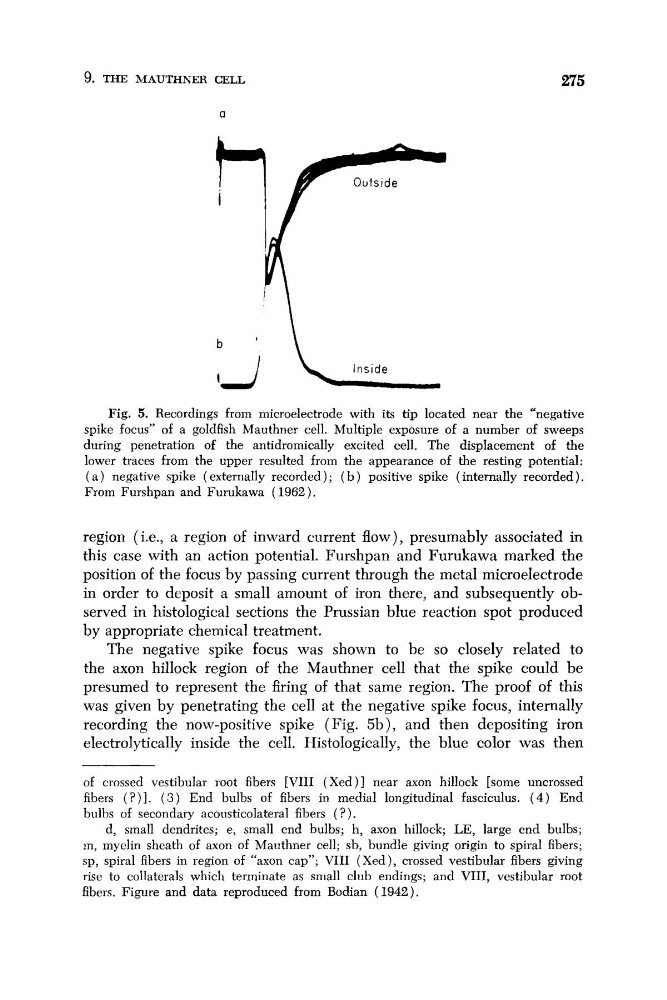
Fig. 5. Recordings from microelectrode with its tip located near the “negative
spike focus” of a goldfish Mauthner cell. Multiple exposure of a number of sweeps
during penetration of the antidromically excited cell. The displacement of the
lower traces from the upper resulted from the appearance of the resting potential:
( a ) negative spike (externally recorded); ( b ) positive spike (internally recorded).
From Furshpan and Furukawa ( 1962).
region (i.e., a region of inward current flow), presumably associated in
this case with an action potential. Furshpan and Furukawa marked the
position of the focus by passing current through the metal microelectrode
in order to deposit a small amount of iron there, and subsequently observed in histological sections the Prussian blue reaction spot produced
by appropriate chemical treatment.
The negative spike focus was shown to be so closely related to
the axon hillock region of the Mauthner cell that the spike could be
presumed to represent the firing of that same region. The proof of this
was given by penetrating the cell at the negative spike focus, internally
recording the now-positive spike (Fig. 5b), and then depositing iron
electrolytically inside the cell. Histologically, the blue color was then
of crossed vestibular root fibers [VIII (Xed)] near axon hillock [some uncrossed
fibers ( ? ) I . ( 3 ) End bulbs of fibers in medial longitudinal fasciculus. ( 4 ) End
bulbs of secondary acousticolateral fibers ( ?).
d, small dendrites; e, small end bulbs; h, axon hillock; LE, large end bulbs;
in, myelin sheath of axon of Manthner cell; sb, bundle giving origin to spiral fibers;
sp, spiral fibers in region of “axon cap”; VIII (Xed), crossed vestibular fibers giving
rise to collaterals which terminate as small club endings; and VIII, vestibular root
fibers. Figure and data reproduced from Bodian (1942).
275
a
b
’
\
Fig. 5. Recordings from microelectrode with its tip located near the “negative
spike focus” of a goldfish Mauthner cell. Multiple exposure of a number of sweeps
during penetration of the antidromically excited cell. The displacement of the
lower traces from the upper resulted from the appearance of the resting potential:
( a ) negative spike (externally recorded); ( b ) positive spike (internally recorded).
From Furshpan and Furukawa ( 1962).
region (i.e., a region of inward current flow), presumably associated in
this case with an action potential. Furshpan and Furukawa marked the
position of the focus by passing current through the metal microelectrode
in order to deposit a small amount of iron there, and subsequently observed in histological sections the Prussian blue reaction spot produced
by appropriate chemical treatment.
The negative spike focus was shown to be so closely related to
the axon hillock region of the Mauthner cell that the spike could be
presumed to represent the firing of that same region. The proof of this
was given by penetrating the cell at the negative spike focus, internally
recording the now-positive spike (Fig. 5b), and then depositing iron
electrolytically inside the cell. Histologically, the blue color was then
of crossed vestibular root fibers [VIII (Xed)] near axon hillock [some uncrossed
fibers ( ? ) I . ( 3 ) End bulbs of fibers in medial longitudinal fasciculus. ( 4 ) End
bulbs of secondary acousticolateral fibers ( ?).
d, small dendrites; e, small end bulbs; h, axon hillock; LE, large end bulbs;
in, myelin sheath of axon of Manthner cell; sb, bundle giving origin to spiral fibers;
sp, spiral fibers in region of “axon cap”; VIII (Xed), crossed vestibular fibers giving
rise to collaterals which terminate as small club endings; and VIII, vestibular root
fibers. Figure and data reproduced from Bodian (1942).