

244
LIKE FLOCK
osmium tetroxide buffered with veronal acetate according to Rhodin
(1954)l was applied in situ with a pipette. The organ was then dissected
free with its bony capsule, rinsed in Ringer, and dehydrated with 70%
alcohol. When appropriate areas had been exposed for scanning microscopy the tissue was frozen in liquid propane at -192°C and freezedried. After mounting with glue on metal holders the specimens were
covered with gold by vacuum evaporation and examined in a Cambridge
Stereoscan microscope. A similar procedure applies to epidermal lateral
line organs which were taken from the salamander, Necturus m u l o s u s .
Specimens for transmission electron microscopy were fixed in osmium
tetroxide, dehydrated in alcohol, embedded in Epon ( Luft, 1961 ) ,
sectioned in an LKB Ultrotome, and examined in a Siemens Elmiskop I.
B. Canal Organs
The sensory area is located at the bottom of a bony half-cylinder
( Fig. 1). At this point the lumen of the canal is narrowed by the presence
of a septum; here the cupula reaches its maximum height and partially
occludes the lumen. If volume displacement occurs between the two
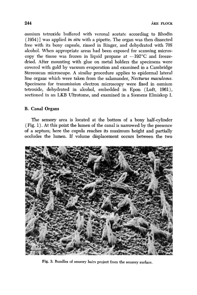
Fig. 3. Bundles of sensory hairs project from the sensory surface.
LIKE FLOCK
osmium tetroxide buffered with veronal acetate according to Rhodin
(1954)l was applied in situ with a pipette. The organ was then dissected
free with its bony capsule, rinsed in Ringer, and dehydrated with 70%
alcohol. When appropriate areas had been exposed for scanning microscopy the tissue was frozen in liquid propane at -192°C and freezedried. After mounting with glue on metal holders the specimens were
covered with gold by vacuum evaporation and examined in a Cambridge
Stereoscan microscope. A similar procedure applies to epidermal lateral
line organs which were taken from the salamander, Necturus m u l o s u s .
Specimens for transmission electron microscopy were fixed in osmium
tetroxide, dehydrated in alcohol, embedded in Epon ( Luft, 1961 ) ,
sectioned in an LKB Ultrotome, and examined in a Siemens Elmiskop I.
B. Canal Organs
The sensory area is located at the bottom of a bony half-cylinder
( Fig. 1). At this point the lumen of the canal is narrowed by the presence
of a septum; here the cupula reaches its maximum height and partially
occludes the lumen. If volume displacement occurs between the two
Fig. 3. Bundles of sensory hairs project from the sensory surface.