

418
JOHAN B. STEEN
v
... , ' .
M 'H 8 C
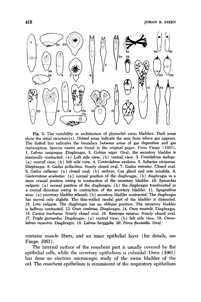
Fig. 3. The variability in architecture of physoclist swim bladders. Dark areas
show the retial structure(s). Dotted areas indicate the area from where gas appears.
The dotted line indicates the boundary between areas of gas deposition and gas
reabsorption. Species names are found in the original paper. From Fange (1953).
1. Labrus ossigragus. Diaphragm. 2. Gobius niger. Oval: the secretory bladder is
maximally contracted. ( a ) Left side view; ( b ) ventral view. 3. Crenilabnrs melops:
( a ) ventral view; ( b ) left side view. 4. Centrolabrus exoletos. 5. Sebastes viviparus.
Diaphragm. 6 . Gadus pollachius. Nearly closed oval. 7. Gadus minutus. Closed oval.
8. Gadus callarius: ( a ) closed oval; ( b ) embryo. Gas gland and rete mirabile. 9.
Gasterosteus aculentus: ( a ) normal position of the diaphragm; ( b ) diaphragm in a
more cranial position owing to contraction of the secretory bladder. 10. Spinachia
vulgaris: ( a ) normal position of the diaphragm; ( b ) the diaphragm translocated in
a cranial direction owing to contraction of the secretory bladder. 11. Syngnathus
acus: ( a ) secretory bladder relaxed; ( b ) secretory bladder contracted. The diaphragm
has moved only slightly. The thin-walled caudal part of the bladder is distended.
12. Lota vulgaris. The diaphragm has an oblique position. The secretory bladder
is halfway contracted, 13. Oms cimbrius. Diaphragm. 14. Onos mustela. Diaphragm.
15. Caranx trachurus. Nearly closed oval. 16. Raniceps raninus. Nearly closed oval.
17. Trigla gurnardus. Diaphragm: ( a ) ventral view; ( b ) left side view. 18. Ctenolabrus rupestris. Diaphragm. 19. Labrus berggylta. 20. Perca fluviatilis. Oval.
contains muscle fibers, and an inner epithelial layer (for details, see
Fange, 1953).
The internal surface of the resorbent part is usually covered by flat
epithelial cells, while the secretory epithelium is cuboidal. Dorn (1961)
has done an electron microscopic study of the swim bladder of the
eel. The resorbent epithelium is reminiscent of the respiratory epithelium
JOHAN B. STEEN
v
... , ' .
M 'H 8 C
Fig. 3. The variability in architecture of physoclist swim bladders. Dark areas
show the retial structure(s). Dotted areas indicate the area from where gas appears.
The dotted line indicates the boundary between areas of gas deposition and gas
reabsorption. Species names are found in the original paper. From Fange (1953).
1. Labrus ossigragus. Diaphragm. 2. Gobius niger. Oval: the secretory bladder is
maximally contracted. ( a ) Left side view; ( b ) ventral view. 3. Crenilabnrs melops:
( a ) ventral view; ( b ) left side view. 4. Centrolabrus exoletos. 5. Sebastes viviparus.
Diaphragm. 6 . Gadus pollachius. Nearly closed oval. 7. Gadus minutus. Closed oval.
8. Gadus callarius: ( a ) closed oval; ( b ) embryo. Gas gland and rete mirabile. 9.
Gasterosteus aculentus: ( a ) normal position of the diaphragm; ( b ) diaphragm in a
more cranial position owing to contraction of the secretory bladder. 10. Spinachia
vulgaris: ( a ) normal position of the diaphragm; ( b ) the diaphragm translocated in
a cranial direction owing to contraction of the secretory bladder. 11. Syngnathus
acus: ( a ) secretory bladder relaxed; ( b ) secretory bladder contracted. The diaphragm
has moved only slightly. The thin-walled caudal part of the bladder is distended.
12. Lota vulgaris. The diaphragm has an oblique position. The secretory bladder
is halfway contracted, 13. Oms cimbrius. Diaphragm. 14. Onos mustela. Diaphragm.
15. Caranx trachurus. Nearly closed oval. 16. Raniceps raninus. Nearly closed oval.
17. Trigla gurnardus. Diaphragm: ( a ) ventral view; ( b ) left side view. 18. Ctenolabrus rupestris. Diaphragm. 19. Labrus berggylta. 20. Perca fluviatilis. Oval.
contains muscle fibers, and an inner epithelial layer (for details, see
Fange, 1953).
The internal surface of the resorbent part is usually covered by flat
epithelial cells, while the secretory epithelium is cuboidal. Dorn (1961)
has done an electron microscopic study of the swim bladder of the
eel. The resorbent epithelium is reminiscent of the respiratory epithelium