

8. THE REGULATION OF BREATHING
315
entrant curves of each branchial arch; there are some smaller muscles
in the interbranchial septa and the gill sacs. The constrictor and diagonal
muscles were shown, by observation and experiments in which electromyograms were recorded, to function together in expiration. No muscles
were active during the inspiration of water, which in the adult lamprey
occurs through the external gill openings. Just as in the dogfish it appears that expansion of the gill chambers is caused by elastic recoil,
although Roberts (1950) noted that this was not a very forceful movement in the cartilaginous branchial skeleton of Lampetru.
Gill ventilation in myxinoids is very different from that described
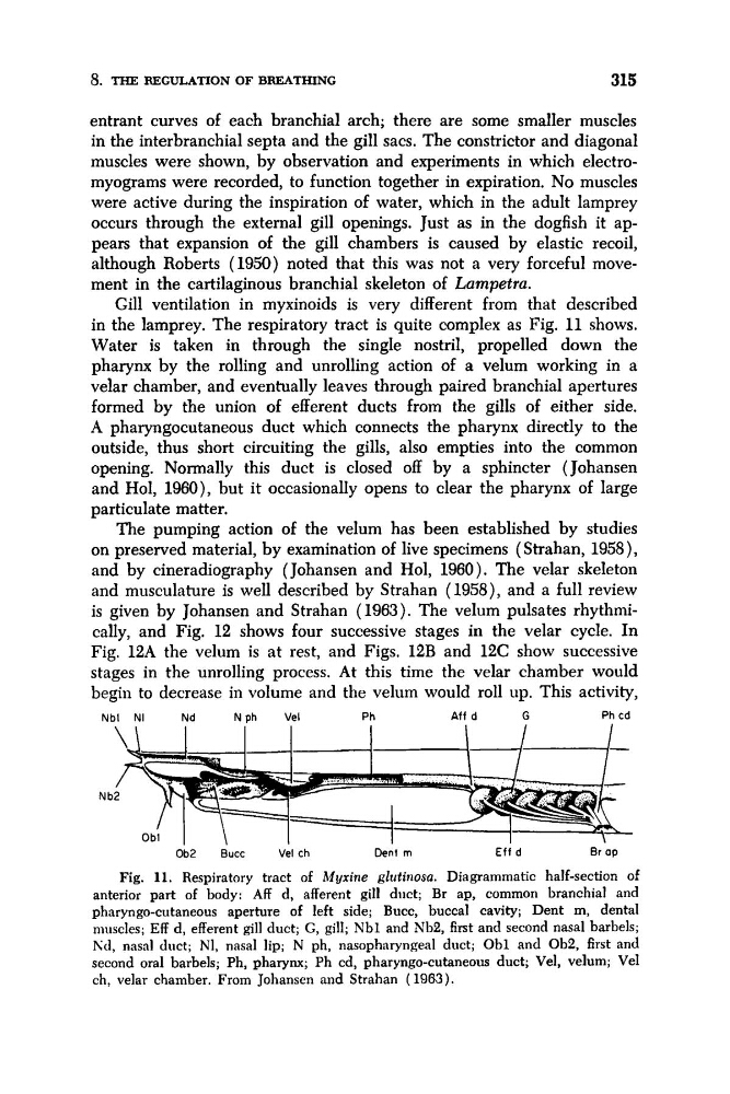
in the lamprey. The respiratory tract is quite complex as Fig. 11 shows.
Water is taken in through the single nostril, propelled down the
pharynx by the rolling and unrolling action of a velum working in a
velar chamber, and eventually leaves through paired branchial apertures
formed by the union of efferent ducts from the gills of either side.
A pharyngocutaneous duct which connects the pharynx directly to the
outside, thus short circuiting the gills, also empties into the common
opening. Normally this duct is closed off by a sphincter (Johansen
and Hol, 1960), but it occasionally opens to clear the pharynx of large
particulate matter.
The pumping action of the velum has been established by studies
on preserved material, by examination of live specimens (Strahan, 1958),
and by cineradiography (Johansen and Hol, 1960). The velar skeleton
and musculature is well described by Strahan (1958), and a full review
is given by Johansen and Strahan (1963). The velum pulsates rhythmically, and Fig. 12 shows four successive stages in the velar cycle. In
Fig. 12A the velum is at rest, and Figs. 12B and 12C show successive
stages in the unrolling process. At this time the velar chamber would
begin to decrease in volume and the velum would roll up. This activity,
Nbl NI
Nd
Nph
Vel
Ph
Aff d
G
Ph c d
N b
Ob2
Bucc
Vel c h
Dent m
Eff d
Br ap
Fig. 11. Respiratory tract of Myrine glutinosa. Diagrammatic half-section of
anterior part of body: Aff d, afferent gill diict; Br ap, common branchial and
pharyngo-cutaneous aperture of left side; Bucc, buccal cavity; Dent m, dental
muscles; Eff d, efferent gill duct; G, gill; Nbl and Nb2, first and second nasal barbels;
Iid, nasal duct; NI, nasal lip; N ph, nasopharyngeal duct; Obl and Ob2, first and
second oral barbels; Ph, pharynx; Ph cd, pharyngo-cutaneous duct; Vel, velum; Vel
ch, velar chamber. From Johansen and Strahan (1963).
315
entrant curves of each branchial arch; there are some smaller muscles
in the interbranchial septa and the gill sacs. The constrictor and diagonal
muscles were shown, by observation and experiments in which electromyograms were recorded, to function together in expiration. No muscles
were active during the inspiration of water, which in the adult lamprey
occurs through the external gill openings. Just as in the dogfish it appears that expansion of the gill chambers is caused by elastic recoil,
although Roberts (1950) noted that this was not a very forceful movement in the cartilaginous branchial skeleton of Lampetru.
Gill ventilation in myxinoids is very different from that described
in the lamprey. The respiratory tract is quite complex as Fig. 11 shows.
Water is taken in through the single nostril, propelled down the
pharynx by the rolling and unrolling action of a velum working in a
velar chamber, and eventually leaves through paired branchial apertures
formed by the union of efferent ducts from the gills of either side.
A pharyngocutaneous duct which connects the pharynx directly to the
outside, thus short circuiting the gills, also empties into the common
opening. Normally this duct is closed off by a sphincter (Johansen
and Hol, 1960), but it occasionally opens to clear the pharynx of large
particulate matter.
The pumping action of the velum has been established by studies
on preserved material, by examination of live specimens (Strahan, 1958),
and by cineradiography (Johansen and Hol, 1960). The velar skeleton
and musculature is well described by Strahan (1958), and a full review
is given by Johansen and Strahan (1963). The velum pulsates rhythmically, and Fig. 12 shows four successive stages in the velar cycle. In
Fig. 12A the velum is at rest, and Figs. 12B and 12C show successive
stages in the unrolling process. At this time the velar chamber would
begin to decrease in volume and the velum would roll up. This activity,
Nbl NI
Nd
Nph
Vel
Ph
Aff d
G
Ph c d
N b
Ob2
Bucc
Vel c h
Dent m
Eff d
Br ap
Fig. 11. Respiratory tract of Myrine glutinosa. Diagrammatic half-section of
anterior part of body: Aff d, afferent gill diict; Br ap, common branchial and
pharyngo-cutaneous aperture of left side; Bucc, buccal cavity; Dent m, dental
muscles; Eff d, efferent gill duct; G, gill; Nbl and Nb2, first and second nasal barbels;
Iid, nasal duct; NI, nasal lip; N ph, nasopharyngeal duct; Obl and Ob2, first and
second oral barbels; Ph, pharynx; Ph cd, pharyngo-cutaneous duct; Vel, velum; Vel
ch, velar chamber. From Johansen and Strahan (1963).