

316
G. SHELTON
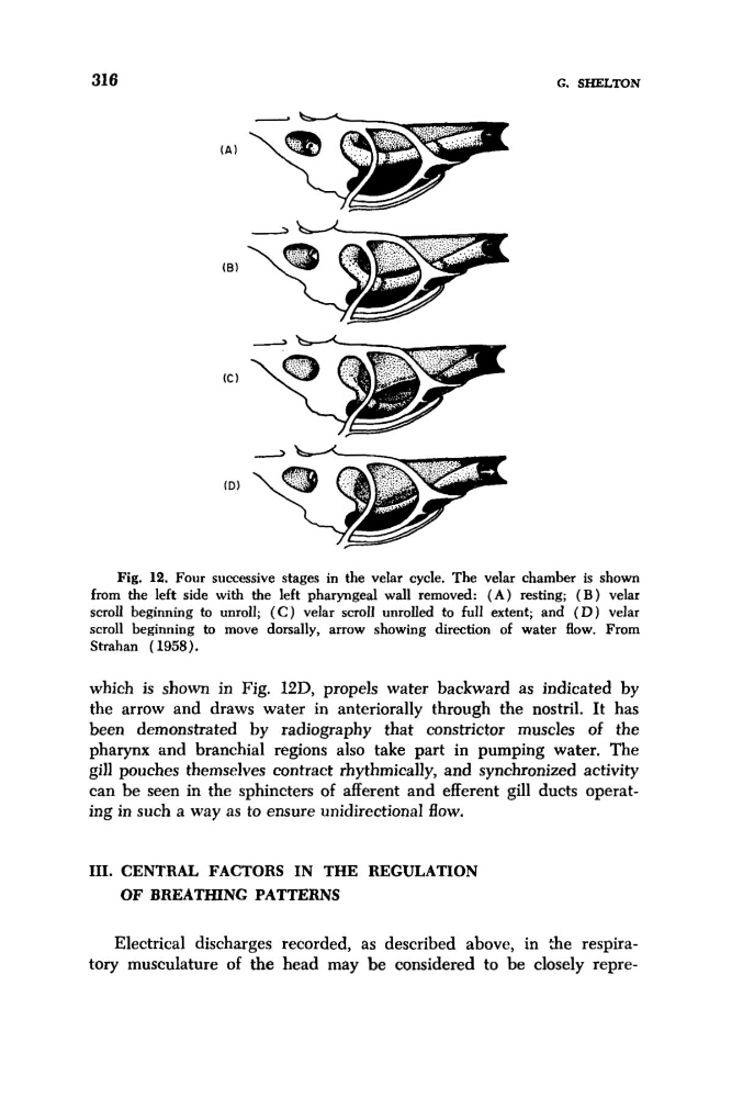
Fig. 12. Four successive stages in the velar cycle. The velar chamber is shown
from the left side with the left pharyngeal wall removed: ( A ) resting; ( B ) velar
scroll beginning to unroll; ( C ) velar scroll unrolled to full extent; and ( D ) velar
scroll beginning to move dorsally, arrow showing direction of water flow. From
Strahan (1958).
which is shown in Fig. l2D, propels water backward as indicated by
the arrow and draws water in anteriorally through the nostril. It has
been demonstrated by radiography that constrictor muscles of the
pharynx and branchial regions also take part in pumping water. The
gill pouches themselves contract rhythmically, and synchronized activity
can be seen in the sphincters of afferent and efferent gill ducts operating in such a way as to ensure unidirectional flow.
111. CENTRAL FACTORS IN THE REGULATION
OF BREATHING PATTERNS
Electrical discharges recorded, as described above, in $e respiratory musculature of the head may be considered to be closely repre-
G. SHELTON
Fig. 12. Four successive stages in the velar cycle. The velar chamber is shown
from the left side with the left pharyngeal wall removed: ( A ) resting; ( B ) velar
scroll beginning to unroll; ( C ) velar scroll unrolled to full extent; and ( D ) velar
scroll beginning to move dorsally, arrow showing direction of water flow. From
Strahan (1958).
which is shown in Fig. l2D, propels water backward as indicated by
the arrow and draws water in anteriorally through the nostril. It has
been demonstrated by radiography that constrictor muscles of the
pharynx and branchial regions also take part in pumping water. The
gill pouches themselves contract rhythmically, and synchronized activity
can be seen in the sphincters of afferent and efferent gill ducts operating in such a way as to ensure unidirectional flow.
111. CENTRAL FACTORS IN THE REGULATION
OF BREATHING PATTERNS
Electrical discharges recorded, as described above, in $e respiratory musculature of the head may be considered to be closely repre-