

5. ACID-BASE BALANCE
197
f
60
PH
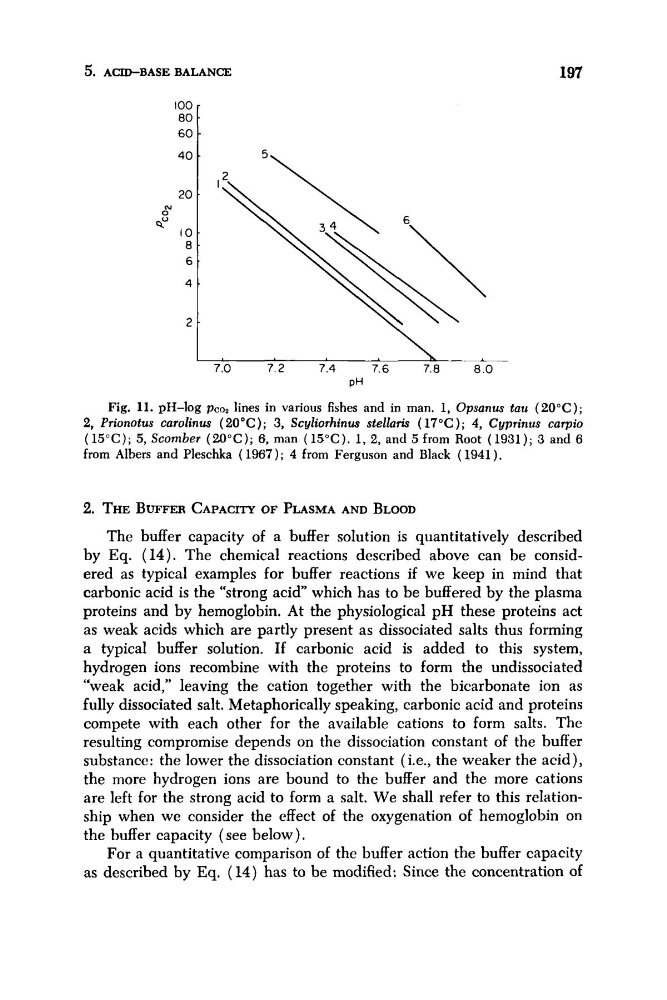
Fig. 11. pH-log pCo2 lines in various fishes and in man. 1, Opsanus tau (20°C);
2, Prionotus carolinus (20°C); 3, Scyliorhinus stellaris (17°C); 4, Cyprinus carpio
(15°C); 5, Scomber (20°C); 6, man (15°C). 1 , 2 , and 5 from Root (1931); 3 and 6
from Albers and Pleschka ( 1967); 4 from Ferguson and Black ( 1941).
2. THE BUFFER CAPACITY OF PLASMA AND BLOOD
The buffer capacity of a buffer solution is quantitatively described
by Eq. (14). The chemical reactions described above can be considered as typical examples for buffer reactions if we keep in mind that
carbonic acid is the “strong a c i d which has to be buffered by the plasma
proteins and by hemoglobin. At the physiological p H these proteins act
as weak acids which are partly present as dissociated salts thus forming
a typical buffer solution. If carbonic acid is added to this system,
hydrogen ions recombine with the proteins to form the undissociated
“weak acid,” leaving the cation together with the bicarbonate ion as
fully dissociated salt. Metaphorically speaking, carbonic acid and proteins
compete with each other for the available cations to form salts. The
resulting compromise depends on the dissociation constant of the buffer
substance: the lower the dissociation constant (i.e., the weaker the acid),
the more hydrogen ions are bound to the buffer and the more cations
are left for the strong acid to form a salt. We shall refer to this relationship when we consider the effect of the oxygenation of hemoglobin on
the buffer capacity (see below).
For a quantitative comparison of the buffer action the buffer capacity
as described by Eq. (14) has to be modified: Since the concentration of
197
f
60
PH
Fig. 11. pH-log pCo2 lines in various fishes and in man. 1, Opsanus tau (20°C);
2, Prionotus carolinus (20°C); 3, Scyliorhinus stellaris (17°C); 4, Cyprinus carpio
(15°C); 5, Scomber (20°C); 6, man (15°C). 1 , 2 , and 5 from Root (1931); 3 and 6
from Albers and Pleschka ( 1967); 4 from Ferguson and Black ( 1941).
2. THE BUFFER CAPACITY OF PLASMA AND BLOOD
The buffer capacity of a buffer solution is quantitatively described
by Eq. (14). The chemical reactions described above can be considered as typical examples for buffer reactions if we keep in mind that
carbonic acid is the “strong a c i d which has to be buffered by the plasma
proteins and by hemoglobin. At the physiological p H these proteins act
as weak acids which are partly present as dissociated salts thus forming
a typical buffer solution. If carbonic acid is added to this system,
hydrogen ions recombine with the proteins to form the undissociated
“weak acid,” leaving the cation together with the bicarbonate ion as
fully dissociated salt. Metaphorically speaking, carbonic acid and proteins
compete with each other for the available cations to form salts. The
resulting compromise depends on the dissociation constant of the buffer
substance: the lower the dissociation constant (i.e., the weaker the acid),
the more hydrogen ions are bound to the buffer and the more cations
are left for the strong acid to form a salt. We shall refer to this relationship when we consider the effect of the oxygenation of hemoglobin on
the buffer capacity (see below).
For a quantitative comparison of the buffer action the buffer capacity
as described by Eq. (14) has to be modified: Since the concentration of