

352
N. BALAKRISHNAN NAIR AND M. SARASWATHY
anterior lobe resembles a triangle which joins the median lobe by one
of its long sides. The free angle of the lobe points forwards like a beak.
At the dorsal free margin, the outline is interrupted by a deep sinus
from which a smooth callus is reflected over its surface. The outer
surface of this lobe is sculptured by fine ridges parallel with the ventral
free margin, extending from the callus on the dorsal margin to the
median lobe, where they meet almost at right angles those borne by the
anterior region of this lobe. The denticulated ridges of the median lobe
run parallel with the anterior free margin, bear stouter denticles and are
more numerous than those on the anterior lobe. Some of the ridges in
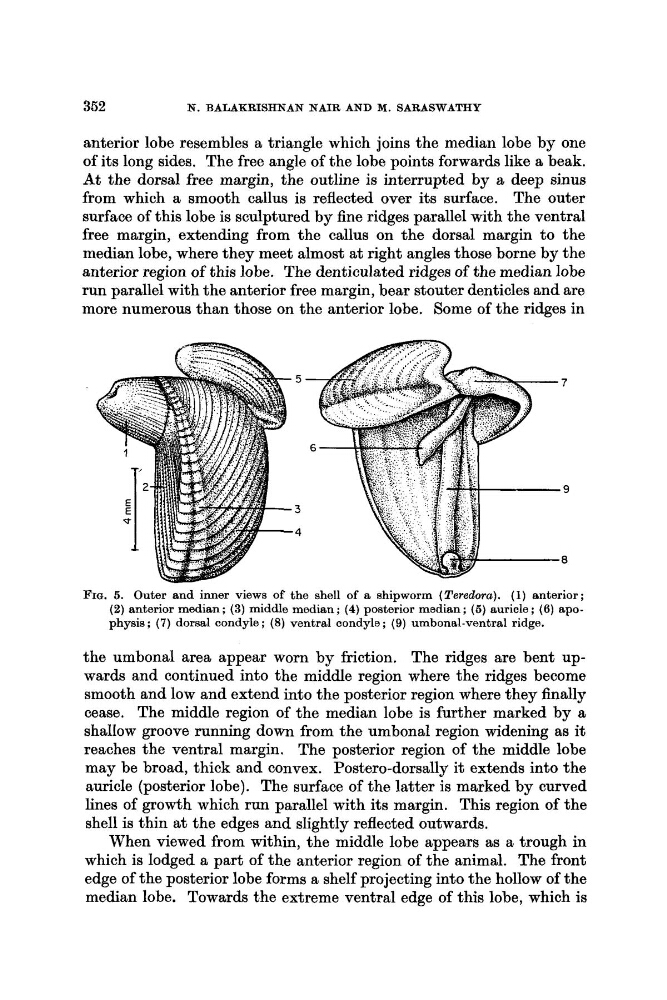
FIG. 5. Outer and inner views of the shell of a shipworm (Teredora). (1) anterior;
(2) anterior median; (3) middle median; (4) posterior median; (5) auricle; (6) apophysis; (7) dorsal condyle ; (8) ventral oondyle ; (9) umbonal-ventral ridge.
the umbonal area appear worn by friction. The ridges are bent upwards and continued into the middle region where the ridges become
smooth and low and extend into the posterior region where they finally
cease. The middle region of the median lobe is further marked by a
shallow groove running down from the umbonal region widening as it
reaches the ventral margin, The posterior region of the middle lobe
may be broad, thick and convex. Postero-dorsally it extends into the
auricle (posterior lobe). The surface of the latter is marked by curved
lines of growth which run parallel with its margin. This region of the
shell is thin at the edges and slightly reflected outwards.
When viewed from within, the middle lobe appears as a trough in
which is lodged a part of the anterior region of the animal. The front
edge of the posterior lobe forms a shelf projecting into the hollow of the
median lobe. Towards the extreme ventral edge of this lobe, which is
N. BALAKRISHNAN NAIR AND M. SARASWATHY
anterior lobe resembles a triangle which joins the median lobe by one
of its long sides. The free angle of the lobe points forwards like a beak.
At the dorsal free margin, the outline is interrupted by a deep sinus
from which a smooth callus is reflected over its surface. The outer
surface of this lobe is sculptured by fine ridges parallel with the ventral
free margin, extending from the callus on the dorsal margin to the
median lobe, where they meet almost at right angles those borne by the
anterior region of this lobe. The denticulated ridges of the median lobe
run parallel with the anterior free margin, bear stouter denticles and are
more numerous than those on the anterior lobe. Some of the ridges in
FIG. 5. Outer and inner views of the shell of a shipworm (Teredora). (1) anterior;
(2) anterior median; (3) middle median; (4) posterior median; (5) auricle; (6) apophysis; (7) dorsal condyle ; (8) ventral oondyle ; (9) umbonal-ventral ridge.
the umbonal area appear worn by friction. The ridges are bent upwards and continued into the middle region where the ridges become
smooth and low and extend into the posterior region where they finally
cease. The middle region of the median lobe is further marked by a
shallow groove running down from the umbonal region widening as it
reaches the ventral margin, The posterior region of the middle lobe
may be broad, thick and convex. Postero-dorsally it extends into the
auricle (posterior lobe). The surface of the latter is marked by curved
lines of growth which run parallel with its margin. This region of the
shell is thin at the edges and slightly reflected outwards.
When viewed from within, the middle lobe appears as a trough in
which is lodged a part of the anterior region of the animal. The front
edge of the posterior lobe forms a shelf projecting into the hollow of the
median lobe. Towards the extreme ventral edge of this lobe, which is