

SOME ASPECTS OF THE BIOLOGY OF THE CHAETOGNATHS
303
Iayer is represented by a thin flat epithelium whose nuclei look somewhat stretched in the direction of the axis of the organ. The opening is,
as we have said, in correspondence with the genital papilla which is a
prominence constituted pre-eminently of glandular cells filled with a
secretion which is strongly eosinophilic.
The abundant material at my disposal has enabled me to make a
detailed study of the development of the ovary of Xpadella. Two
groups of cellular elements contribute to the formation of the ovary:
all the oogonia derive from the first two gonocytes which in the larva
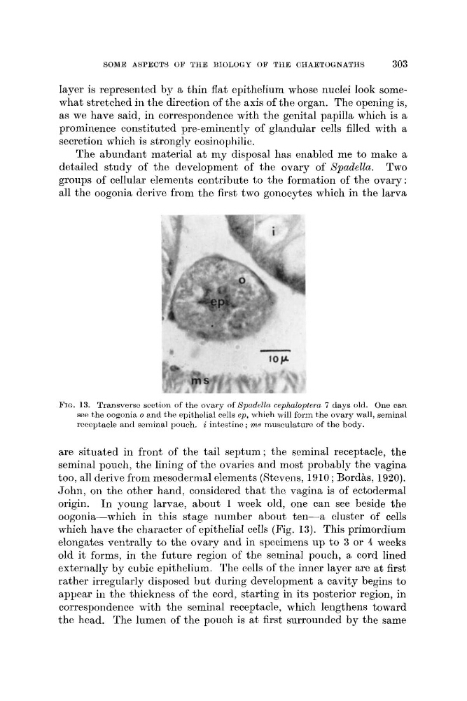
FIG. 13. Transverso soction of the ovary of Spudella cephaloptera 7 days old. One can
see the oogonia o anti the epithelial cells ep, which will form the ovary wall, seminal
receptacle and seminal pourh. i intestine ; 'm,,s musculature of the body.
are situated in front of the tail septum ; the seminal receptacle, the
seminal pouch, the lining of the ovaries and most probably the vagina
too, all derive from mesodermal elements (Etevens, 1910 ; Bordhs, 1920).
John, on the other hand, considered that the vagina is of ectodermal
origin. In young larvae, about 1 week old, one can see beside the
oogonia-which in this stage number about ten-a cluster of cells
which have the character of epithelial cells (Fig. 13). This primordium
elongates ventrally to the ovary and in specimens up to 3 or 4 weeks
old it forms, in the future region of the seminal pouch, a cord lined
externally by cubic epithelium. The cells of the inner layer are at first
rather irregularly disposed but during development a cavity begins to
appear in the thickness of the cord, starting in its posterior region, in
correspondence with the seminal receptacle, which lengthens toward
the head. The lumen of the pouch is at first surrounded by the same
303
Iayer is represented by a thin flat epithelium whose nuclei look somewhat stretched in the direction of the axis of the organ. The opening is,
as we have said, in correspondence with the genital papilla which is a
prominence constituted pre-eminently of glandular cells filled with a
secretion which is strongly eosinophilic.
The abundant material at my disposal has enabled me to make a
detailed study of the development of the ovary of Xpadella. Two
groups of cellular elements contribute to the formation of the ovary:
all the oogonia derive from the first two gonocytes which in the larva
FIG. 13. Transverso soction of the ovary of Spudella cephaloptera 7 days old. One can
see the oogonia o anti the epithelial cells ep, which will form the ovary wall, seminal
receptacle and seminal pourh. i intestine ; 'm,,s musculature of the body.
are situated in front of the tail septum ; the seminal receptacle, the
seminal pouch, the lining of the ovaries and most probably the vagina
too, all derive from mesodermal elements (Etevens, 1910 ; Bordhs, 1920).
John, on the other hand, considered that the vagina is of ectodermal
origin. In young larvae, about 1 week old, one can see beside the
oogonia-which in this stage number about ten-a cluster of cells
which have the character of epithelial cells (Fig. 13). This primordium
elongates ventrally to the ovary and in specimens up to 3 or 4 weeks
old it forms, in the future region of the seminal pouch, a cord lined
externally by cubic epithelium. The cells of the inner layer are at first
rather irregularly disposed but during development a cavity begins to
appear in the thickness of the cord, starting in its posterior region, in
correspondence with the seminal receptacle, which lengthens toward
the head. The lumen of the pouch is at first surrounded by the same