

301
ELVEZIO GHIRARDELLI
cells of the cord which later form the syncytium and which will later
completely line the pouch and the receptacle.
While the pouch and the seminal receptacle are developing, the
oogonia have multiplied and the maturation prophase has begun. The
young oocytes at the anterior end of the ovary do not always appear
to be in contact with the walls of the seminal pouch ; when, however,
they have reached a certain development, one sees that the two cells
of the outer epithelial layer of the pouch become more elongated,
assuming therefore somewhat larger dimensions than the nearby cells.
Their appearance is often like that of a kidney or a bean so that the
two cells, one in front of the other, look a little like a stoma of the leaves.
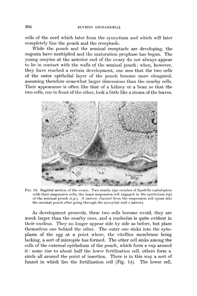
FIG. 14. Sagittal section of the ovary. Two nearly ripe oocytes of SpadPlZa cephabptera
with their suspension cells, the inner suspension cell engaged in the epithclium (ep)
of the seminal pouch (s.P.). A narrow channel from the suspension cell opens into
the seminal pouch after going through the syncgtial wall s (arrow).
As development proceeds, these two cells become ovoid, they are
much larger than the nearby ones, and a nucleolus is quite evident in
their nucleus. They no longer appear side by side as before, but place
themselves one behind the other. The outer one sinks into the cytoplasm of the egg at a point where, the vitelline membrane being
lacking, a sort of micropile has formed. The other cell sinks among the
cells of the external epithelium of the pouch, which form a cup around
it : some rise to about half the lower fertilization cell, others form a
circle all around the point of insertion. There is in this way a sort of
funnel in which lies the fertilization cell (Fig. 14). The lower cell,
ELVEZIO GHIRARDELLI
cells of the cord which later form the syncytium and which will later
completely line the pouch and the receptacle.
While the pouch and the seminal receptacle are developing, the
oogonia have multiplied and the maturation prophase has begun. The
young oocytes at the anterior end of the ovary do not always appear
to be in contact with the walls of the seminal pouch ; when, however,
they have reached a certain development, one sees that the two cells
of the outer epithelial layer of the pouch become more elongated,
assuming therefore somewhat larger dimensions than the nearby cells.
Their appearance is often like that of a kidney or a bean so that the
two cells, one in front of the other, look a little like a stoma of the leaves.
FIG. 14. Sagittal section of the ovary. Two nearly ripe oocytes of SpadPlZa cephabptera
with their suspension cells, the inner suspension cell engaged in the epithclium (ep)
of the seminal pouch (s.P.). A narrow channel from the suspension cell opens into
the seminal pouch after going through the syncgtial wall s (arrow).
As development proceeds, these two cells become ovoid, they are
much larger than the nearby ones, and a nucleolus is quite evident in
their nucleus. They no longer appear side by side as before, but place
themselves one behind the other. The outer one sinks into the cytoplasm of the egg at a point where, the vitelline membrane being
lacking, a sort of micropile has formed. The other cell sinks among the
cells of the external epithelium of the pouch, which form a cup around
it : some rise to about half the lower fertilization cell, others form a
circle all around the point of insertion. There is in this way a sort of
funnel in which lies the fertilization cell (Fig. 14). The lower cell,