

302
ELVEZIO GIIIRANDELLI
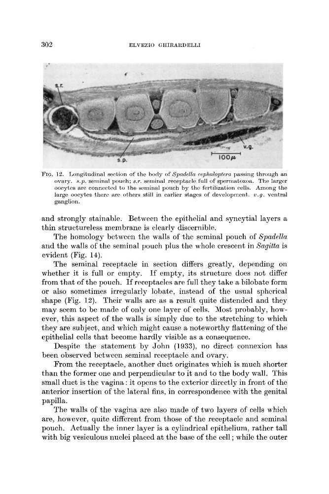
FIG. 12. Longitudinal section of the body of Spadella cephaloptero passing through an
ovary. s.p. seminal pouch; s.r. seminal receptacle full of spermatozoa. The larger
oocytes are connccted t o the scrninal pouch by the fertilization cells. Among the
large oocytes thero are others still in earlier stages of development. w.g. ventral
ganglion.
and strongly stainable. Between the epithelial and syncytial layers a
thin structureless membrane is clearly discernible.
The homology between the walls of the seminal pouch of Spadella
and the walls of the seminal pouch plus the whole crescent in Xagitfa is
evident (Fig. 14).
The seminal receptacle in section differs greatly, depending on
whether it is full or empty. If empty, its structure does not differ
from that of the pouch. If receptacles are full they take a bilobate form
or also sometimes irregularly lobate, instead of the usual sphcrical
shape (Fig. 12). Their walls are as a result quite distended and they
may seem to be made of only one layer of cells. Most probably, however, this aspect of the walls is simply due to the stretching to which
they are subject, and which might cause a noteworthy flattening of the
epithelial cells that become hardly visible as a consequence.
Despite the statement by John (1933), no direct connexion has
been observed between seminal receptacle and ovary.
From the receptacle, another duct originates which is much shorter
than the former one and perpendicular to it and to the body wall. This
small duct is the vagina : it opens to the exterior directly in front of the
anterior insertion of the lateral fins, in correspondence with the genital
papilla.
The walls of the vagina are also made of two layers of cells w-hich
are, however, quite different from those of the receptacle and seminal
pouch. Actually the inner layer is a cylindrical epithelium, rather tall
with big vesiculous nuclei placed a t the base of the cell ; while the outer
ELVEZIO GIIIRANDELLI
FIG. 12. Longitudinal section of the body of Spadella cephaloptero passing through an
ovary. s.p. seminal pouch; s.r. seminal receptacle full of spermatozoa. The larger
oocytes are connccted t o the scrninal pouch by the fertilization cells. Among the
large oocytes thero are others still in earlier stages of development. w.g. ventral
ganglion.
and strongly stainable. Between the epithelial and syncytial layers a
thin structureless membrane is clearly discernible.
The homology between the walls of the seminal pouch of Spadella
and the walls of the seminal pouch plus the whole crescent in Xagitfa is
evident (Fig. 14).
The seminal receptacle in section differs greatly, depending on
whether it is full or empty. If empty, its structure does not differ
from that of the pouch. If receptacles are full they take a bilobate form
or also sometimes irregularly lobate, instead of the usual sphcrical
shape (Fig. 12). Their walls are as a result quite distended and they
may seem to be made of only one layer of cells. Most probably, however, this aspect of the walls is simply due to the stretching to which
they are subject, and which might cause a noteworthy flattening of the
epithelial cells that become hardly visible as a consequence.
Despite the statement by John (1933), no direct connexion has
been observed between seminal receptacle and ovary.
From the receptacle, another duct originates which is much shorter
than the former one and perpendicular to it and to the body wall. This
small duct is the vagina : it opens to the exterior directly in front of the
anterior insertion of the lateral fins, in correspondence with the genital
papilla.
The walls of the vagina are also made of two layers of cells w-hich
are, however, quite different from those of the receptacle and seminal
pouch. Actually the inner layer is a cylindrical epithelium, rather tall
with big vesiculous nuclei placed a t the base of the cell ; while the outer