

12. QUINONES: STRUCTURE AND DISTRIBUTION
691
quinones. Dimelow (263) has isolated a very small amount of a red
pigment occurring in the eggs and connective tissue of the macrophreate
species Antedon biftda (Pennant). It gives a violet solution in aqueous
sodium bicarbonate, and spectroscopically it could be either a hydroxynaphthoquinone or a hydroxyanthraquinone, but its high acidity (pK
approximately 3.8) favors the former (264). Sutherland and Wells
(265) have recently obtained comparatively large quantities of pigment
from the Australian oligophreate species ComatvL· pectinata Linn,
(about 5% of the dry weight). Chromatography revealed a mixture of
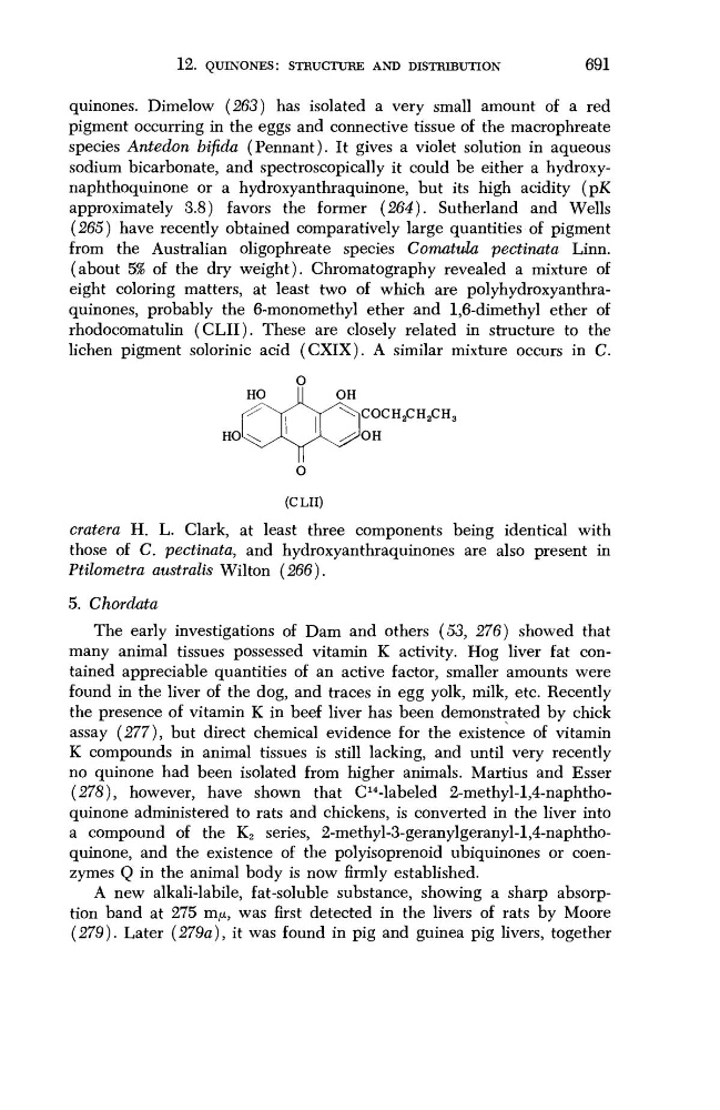
eight coloring matters, at least two of which are polyhydroxyanthraquinones, probably the 6-monomethyl ether and 1,6-dimethyl ether of
rhodocomatulin (CLII). These are closely related in structure to the
lichen pigment solorinic acid (CXIX). A similar mixture occurs in C.
OH
< ^COCH 2 CH 2 CH 3
^OH
O
(CLII)
cratera H. L. Clark, at least three components being identical with
those of C. pectinata, and hydroxyanthraquinones are also present in
Ptilometra australis Wilton (266).
5. Chordata
The early investigations of Dam and others (53, 276) showed that
many animal tissues possessed vitamin K activity. Hog liver fat contained appreciable quantities of an active factor, smaller amounts were
found in the liver of the dog, and traces in egg yolk, milk, etc. Recently
the presence of vitamin K in beef liver has been demonstrated by chick
assay (277), but direct chemical evidence for the existence of vitamin
K compounds in animal tissues is still lacking, and until very recently
no quinone had been isolated from higher animals. Martius and Esser
(278), however, have shown that C
14 -labeled 2-methyl-l,4-naphthoquinone administered to rats and chickens, is converted in the liver into
a compound of the K 2 series, 2-methyl-3-geranylgeranyl-l,4-naphthoquinone, and the existence of the polyisoprenoid ubiquinones or coenzymes Q in the animal body is now firmly established.
A new alkali-labile, fat-soluble substance, showing a sharp absorption band at 275 m/x, was first detected in the livers of rats by Moore
(279). Later (279a), it was found in pig and guinea pig livers, together