

H 2 C *
6 C
CH 3
H
CH 3
H
CH 3
I
I
I
!
I
5 > / 7 XT9 \(/ΐΐ\/ 13MX15
I
I
I
I
H
H
H
H
H 2 C s 2 1 C—CH 3
all-£rcms Vitamin A
Retinene
I
H
CH 3
H
CH 8
H
CH 3
H
I
I
I
.
H r /
c
\/
c
v/
c
v/
c
v/
c
\c/
H 2 C
C
|
|
|
|
H
H
H
CH 2 OH
13-CTS (neo-a)
H2C
C—CH 3
\ c /l
I CH 3
H 2
CH 3
H
CH 3
I
I
I
H
H 2 c/
C
\c/
C
\c/
C
V/
9-cis (iso-a)
H
C
H
H 2 C
C—CH 3
/ χ>0^
*YU
H
1 H
CH 2 OH
CH 3
H
CH 3
I
I
I
H
H
H
H 2 C
C—CH 3
/C v
/
9,13-dicis (iso-b)
XK|
H '
X( f
H 2
CH °
/
C \ C /
CH 3
I
H
CH 3
H
CH 3
H
CH 2 OH
I
I
I
/C^
/C^
/C^
/C*
H 2 C
X ^C^ ^C^ Vc/ ^C^
U-cis (neo-b)
H
H
H
I
H 2 C
C—CH 3
/C
■CX /.„
CH 3
I
H 2
CH 3
CH 2 OH
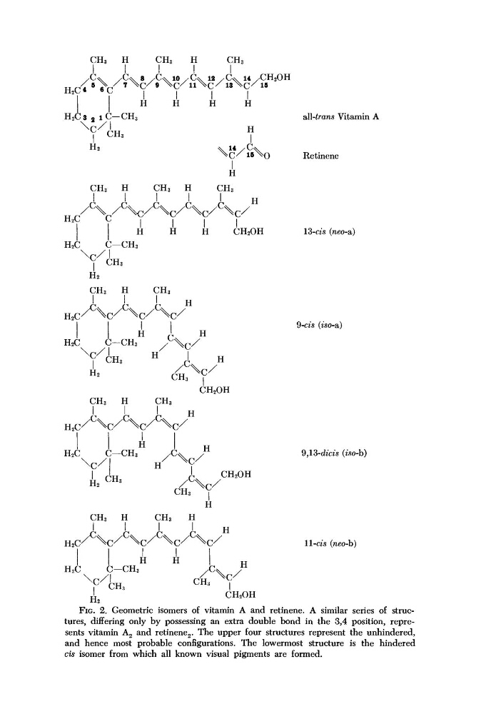
FIG. 2. Geometric isomers of vitamin A and retinene. A similar series of structures, differing only by possessing an extra double bond in the 3,4 position, represents vitamin A 2 and retinene 2 . The upper four structures represent the unhindered,
and hence most probable configurations. The lowermost structure is the hindered
eis isomer from which all known visual pigments are formed.
6 C
CH 3
H
CH 3
H
CH 3
I
I
I
!
I
5 > / 7 XT9 \(/ΐΐ\/ 13MX15
I
I
I
I
H
H
H
H
H 2 C s 2 1 C—CH 3
all-£rcms Vitamin A
Retinene
I
H
CH 3
H
CH 8
H
CH 3
H
I
I
I
.
H r /
c
\/
c
v/
c
v/
c
v/
c
\c/
H 2 C
C
|
|
|
|
H
H
H
CH 2 OH
13-CTS (neo-a)
H2C
C—CH 3
\ c /l
I CH 3
H 2
CH 3
H
CH 3
I
I
I
H
H 2 c/
C
\c/
C
\c/
C
V/
9-cis (iso-a)
H
C
H
H 2 C
C—CH 3
/ χ>0^
*YU
H
1 H
CH 2 OH
CH 3
H
CH 3
I
I
I
H
H
H
H 2 C
C—CH 3
/C v
/
9,13-dicis (iso-b)
XK|
H '
X( f
H 2
CH °
/
C \ C /
CH 3
I
H
CH 3
H
CH 3
H
CH 2 OH
I
I
I
/C^
/C^
/C^
/C*
H 2 C
X ^C^ ^C^ Vc/ ^C^
U-cis (neo-b)
H
H
H
I
H 2 C
C—CH 3
/C
■CX /.„
CH 3
I
H 2
CH 3
CH 2 OH
FIG. 2. Geometric isomers of vitamin A and retinene. A similar series of structures, differing only by possessing an extra double bond in the 3,4 position, represents vitamin A 2 and retinene 2 . The upper four structures represent the unhindered,
and hence most probable configurations. The lowermost structure is the hindered
eis isomer from which all known visual pigments are formed.