

6. PHOTOTROPISM AND PHOTOTAXIS
277
tor the photoreceptor, and their idea was extended, specifically to a
carotenoid, by Voerkel (102). The two peaks in the action spectrum
supported the idea of a carotenoid, and Wald and duBuy (103) found
carotenoid pigments in the light sensitive zones of Avena. In hexane
250
300
350
400
450
500
Wavelength in π\μ
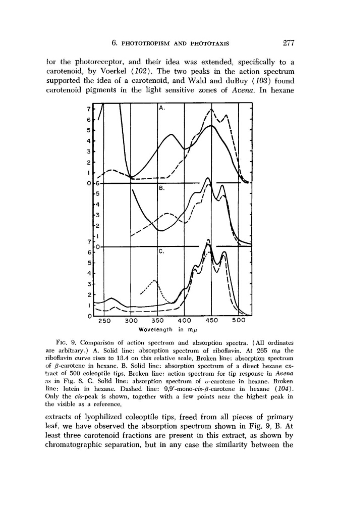
FIG. 9. Comparison of action spectrum and absorption spectra. (All ordinates
are arbitrary.) A. Solid line: absorption spectrum of riboflavin. At 265 m/x the
riboflavin curve rises to 13.4 on this relative scale. Broken line: absorption spectrum
of /^-carotene in hexane. B. Solid line: absorption spectrum of a direct hexane extract of 500 coleoptile tips. Broken line: action spectrum for tip response in Avena
as in Fig. 8. C. Solid line: absorption spectrum of α-carotene in hexane. Broken
line: lutein in hexane. Dashed line: 9,9'-mono-cis-/?-carotene in hexane
(104).
Only the cis-peak is shown, together with a few points near the highest peak in
the visible as a reference.
extracts of lyophilized coleoptile tips, freed from all pieces of primary
leaf, we have observed the absorption spectrum shown in Fig. 9, B. At
least three carotenoid fractions are present in this extract, as shown by
Chromatographie separation, but in any case the similarity between the
277
tor the photoreceptor, and their idea was extended, specifically to a
carotenoid, by Voerkel (102). The two peaks in the action spectrum
supported the idea of a carotenoid, and Wald and duBuy (103) found
carotenoid pigments in the light sensitive zones of Avena. In hexane
250
300
350
400
450
500
Wavelength in π\μ
FIG. 9. Comparison of action spectrum and absorption spectra. (All ordinates
are arbitrary.) A. Solid line: absorption spectrum of riboflavin. At 265 m/x the
riboflavin curve rises to 13.4 on this relative scale. Broken line: absorption spectrum
of /^-carotene in hexane. B. Solid line: absorption spectrum of a direct hexane extract of 500 coleoptile tips. Broken line: action spectrum for tip response in Avena
as in Fig. 8. C. Solid line: absorption spectrum of α-carotene in hexane. Broken
line: lutein in hexane. Dashed line: 9,9'-mono-cis-/?-carotene in hexane
(104).
Only the cis-peak is shown, together with a few points near the highest peak in
the visible as a reference.
extracts of lyophilized coleoptile tips, freed from all pieces of primary
leaf, we have observed the absorption spectrum shown in Fig. 9, B. At
least three carotenoid fractions are present in this extract, as shown by
Chromatographie separation, but in any case the similarity between the