

U L T R A S T R U C T U R E OF T H E A M P H I B I A N EGG
183
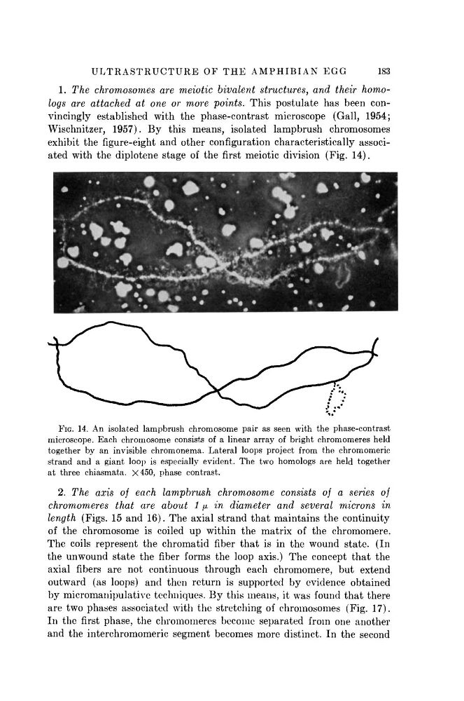
1. The chromosomes are meiotic bivalent structures, and their homologs are attached at one or more points. This postulate has been convincingly established with the phase-contrast microscope (Gall, 1954;
Wischnitzer, 1957). B y this means, isolated lampbrush chromosomes
exhibit the figure-eight and other configuration characteristically associated with the diplotene stage of the first meiotic division (Fig. 14).
FIG. 14. An isolated lampbrush chromosome pair as seen with the phase-contrast
microscope. Each chromosome consists of a linear array of bright chromomeres held
together by an invisible chromonema. Lateral loops project from the chromomeric
strand and a giant loop is especially evident. The two homologs are held together
at three chiasmata. X450, phase contrast.
2. The axis of each lampbrush chromosome consists of a series of
chromomeres that are about 1 μ in diameter and several microns in
length (Figs. 15 and 16). The axial strand that maintains the continuity
of the chromosome is coiled up within the matrix of the chromomere.
The coils represent the chromatid fiber that is in the wound state. (In
the unwound state the fiber forms the loop axis.) The concept that the
axial fibers are not continuous through each chromomere, but extend
outward (as loops) and then return is supported by evidence obtained
by micromanipulative techniques. By this means, it was found that there
are two phases associated with the stretching of chromosomes (Fig. 17).
In the first phase, the chromomeres become separated from one another
and the interchromomeric segment becomes more distinct. In the second
183
1. The chromosomes are meiotic bivalent structures, and their homologs are attached at one or more points. This postulate has been convincingly established with the phase-contrast microscope (Gall, 1954;
Wischnitzer, 1957). B y this means, isolated lampbrush chromosomes
exhibit the figure-eight and other configuration characteristically associated with the diplotene stage of the first meiotic division (Fig. 14).
FIG. 14. An isolated lampbrush chromosome pair as seen with the phase-contrast
microscope. Each chromosome consists of a linear array of bright chromomeres held
together by an invisible chromonema. Lateral loops project from the chromomeric
strand and a giant loop is especially evident. The two homologs are held together
at three chiasmata. X450, phase contrast.
2. The axis of each lampbrush chromosome consists of a series of
chromomeres that are about 1 μ in diameter and several microns in
length (Figs. 15 and 16). The axial strand that maintains the continuity
of the chromosome is coiled up within the matrix of the chromomere.
The coils represent the chromatid fiber that is in the wound state. (In
the unwound state the fiber forms the loop axis.) The concept that the
axial fibers are not continuous through each chromomere, but extend
outward (as loops) and then return is supported by evidence obtained
by micromanipulative techniques. By this means, it was found that there
are two phases associated with the stretching of chromosomes (Fig. 17).
In the first phase, the chromomeres become separated from one another
and the interchromomeric segment becomes more distinct. In the second