

U L T R A S T R U C T U R E OF T H E A M P H I B I A N EGG
175
Rouiller, 1957; Wischnitzer, 1958; Wartenberg, 1962). These masses (as
well as the annuli) are seen in osmium-fixed tissues but not in those
preserved by KMn0 4 (Gall, 1959; Merriam, 1961; Wartenberg, 1962).
It was shown that the different configurations exhibited by these masses
in perpendicular section, could best be interpreted as tubes or cylinders
(Afzelius, 1955; Wischnitzer, 1958). These tubes, corresponding to the
annuli that are seen in oblique or tangential sections of the nuclear
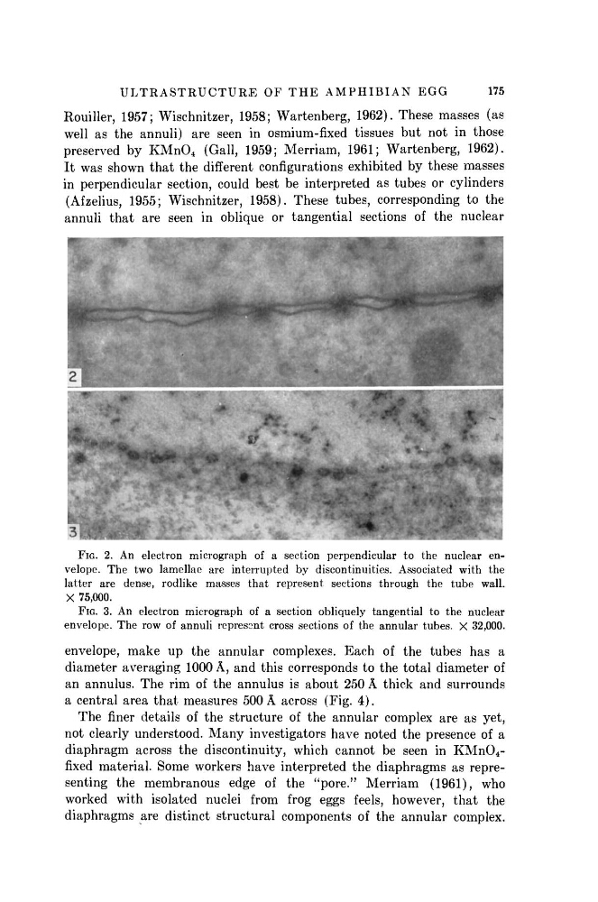
FIG. 2. An electron micrograph of a section perpendicular to the nuclear envelope. The two lamellae are interrupted by discontinuities. Associated with the
latter are dense, rodlike masses that represent sections through the tube wall.
X 75,000.
FIG. 3. An electron micrograph of a section obliquely tangential to the nuclear
envelope. The row of annuli represent cross sections of the annular tubes. X 32,000.
envelope, make up the annular complexes. Each of the tubes has a
diameter averaging 1000 Â, and this corresponds to the total diameter of
an annulus. The rim of the annulus is about 250 Â thick and surrounds
a central area that measures 500 Â across (Fig. 4).
The finer details of the structure of the annular complex are as yet,
not clearly understood. Many investigators have noted the presence of a
diaphragm across the discontinuity, which cannot be seen in KMn0 4 -
fixed material. Some workers have interpreted the diaphragms as representing the membranous edge of the "pore." Merriam (1961), who
worked with isolated nuclei from frog eggs feels, however, that the
diaphragms are distinct structural components of the annular complex.
175
Rouiller, 1957; Wischnitzer, 1958; Wartenberg, 1962). These masses (as
well as the annuli) are seen in osmium-fixed tissues but not in those
preserved by KMn0 4 (Gall, 1959; Merriam, 1961; Wartenberg, 1962).
It was shown that the different configurations exhibited by these masses
in perpendicular section, could best be interpreted as tubes or cylinders
(Afzelius, 1955; Wischnitzer, 1958). These tubes, corresponding to the
annuli that are seen in oblique or tangential sections of the nuclear
FIG. 2. An electron micrograph of a section perpendicular to the nuclear envelope. The two lamellae are interrupted by discontinuities. Associated with the
latter are dense, rodlike masses that represent sections through the tube wall.
X 75,000.
FIG. 3. An electron micrograph of a section obliquely tangential to the nuclear
envelope. The row of annuli represent cross sections of the annular tubes. X 32,000.
envelope, make up the annular complexes. Each of the tubes has a
diameter averaging 1000 Â, and this corresponds to the total diameter of
an annulus. The rim of the annulus is about 250 Â thick and surrounds
a central area that measures 500 Â across (Fig. 4).
The finer details of the structure of the annular complex are as yet,
not clearly understood. Many investigators have noted the presence of a
diaphragm across the discontinuity, which cannot be seen in KMn0 4 -
fixed material. Some workers have interpreted the diaphragms as representing the membranous edge of the "pore." Merriam (1961), who
worked with isolated nuclei from frog eggs feels, however, that the
diaphragms are distinct structural components of the annular complex.