

SAUL W I S C H N I T Z E R
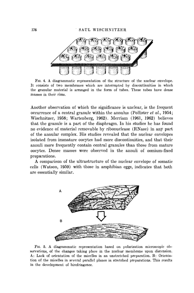
FIG. 4. A diagrammatic representation of the structure of the nuclear envelope.
It consists of two membranes which are interrupted by discontinuities in which
the granular material is arranged in the form of tubes. These tubes have dense
masses in their rims.
Another observation of which the significance is unclear, is the frequent
occurrence of a central granule within the annulus (Pollister et al., 1954;
Wischnitzer, 1958; Wartenberg, 1962). Merriam (1961, 1962) believes
that the granule is a part of the diaphragm. In his studies he has found
no evidence of material removable by ribonuclease (RNase) in any part
of the annular complex. His studies revealed that the nuclear envelopes
isolated from immature oocytes had more discontinuities, and that their
annuli more frequently contain central granules than those from mature
oocytes. Dense masses were observed in the annuli of osmium-fixed
preparations.
A comparison of the ultrastructure of the nuclear envelope of somatic
cells (Watson, 1959) with those in amphibian eggs, indicates that both
are essentially similar.
FIG. 5. A diagrammatic representation based on polarization microscopic observations, of the changes taking place in the nuclear membrane upon distension.
A: Lack of orientation of the micelles in an unstretched preparation. B: Orientation of the micelles in several parallel planes in stretched preparations. This results
in the development of birefringence.
176
FIG. 4. A diagrammatic representation of the structure of the nuclear envelope.
It consists of two membranes which are interrupted by discontinuities in which
the granular material is arranged in the form of tubes. These tubes have dense
masses in their rims.
Another observation of which the significance is unclear, is the frequent
occurrence of a central granule within the annulus (Pollister et al., 1954;
Wischnitzer, 1958; Wartenberg, 1962). Merriam (1961, 1962) believes
that the granule is a part of the diaphragm. In his studies he has found
no evidence of material removable by ribonuclease (RNase) in any part
of the annular complex. His studies revealed that the nuclear envelopes
isolated from immature oocytes had more discontinuities, and that their
annuli more frequently contain central granules than those from mature
oocytes. Dense masses were observed in the annuli of osmium-fixed
preparations.
A comparison of the ultrastructure of the nuclear envelope of somatic
cells (Watson, 1959) with those in amphibian eggs, indicates that both
are essentially similar.
FIG. 5. A diagrammatic representation based on polarization microscopic observations, of the changes taking place in the nuclear membrane upon distension.
A: Lack of orientation of the micelles in an unstretched preparation. B: Orientation of the micelles in several parallel planes in stretched preparations. This results
in the development of birefringence.
176