

V I T E L L I N E M E M B R A N E A N D C O R T I C A L P A R T I C L E S 2 9 5
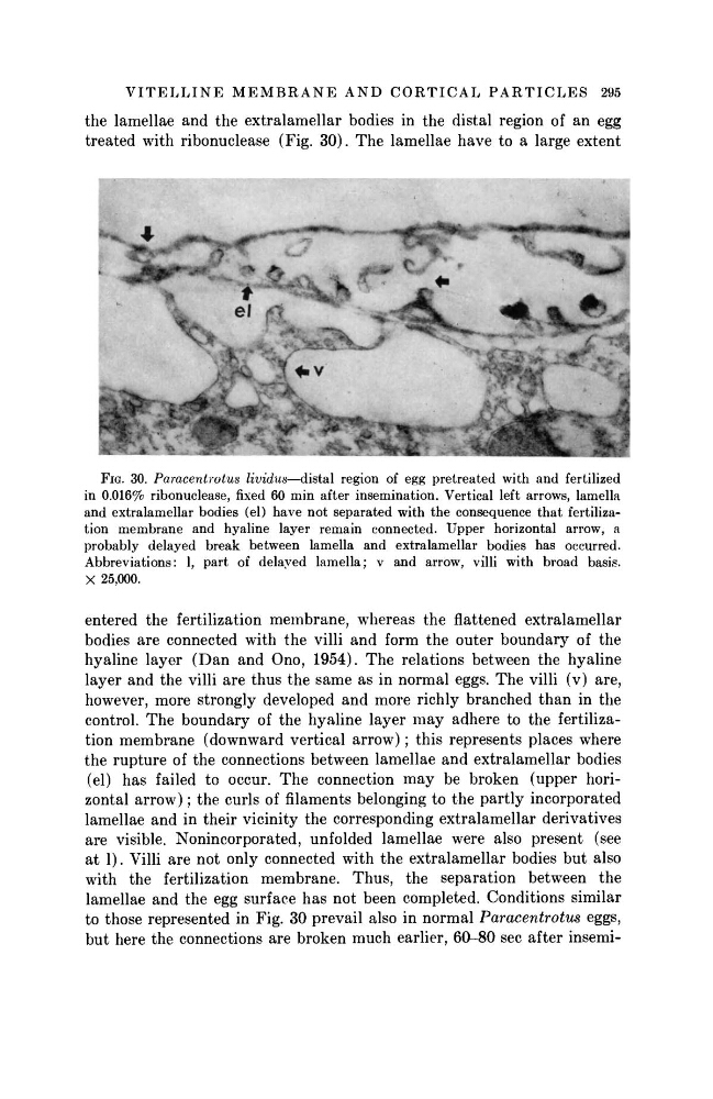
FIG. 3 0 . Paracentrotus lividus—distal region of egg pretreated with and fertilized
in 0.016% ribonuclease, fixed 6 0 min after insemination. Vertical left arrows, lamella
and extralamellar bodies (el) have not separated with the consequence that fertilization membrane and hyaline layer remain connected. Upper horizontal arrow, a
probably delayed break between lamella and extralamellar bodies has occurred.
Abbreviations: 1, part of delayed lamella; ν and arrow, villi with broad basis.
X 25,000.
entered the fertilization membrane, whereas the flattened extralamellar
bodies are connected with the villi and form the outer boundary of the
hyaline layer ( D a n and Ono, 1954). The relations between the hyaline
layer and the villi are thus the same as in normal eggs. The villi (v) are,
however, more strongly developed and more richly branched t h a n in the
control. The boundary of the hyaline layer m a y adhere to the fertilization membrane (downward vertical arrow) ; this represents places where
the rupture of the connections between lamellae and extralamellar bodies
(el) has failed to occur. The connection m a y be broken (upper horizontal arrow) ; the curls of filaments belonging to the partly incorporated
lamellae and in their vicinity the corresponding extralamellar derivatives
are visible. Nonincorporated, unfolded lamellae were also present (see
at 1). Villi are not only connected with the extralamellar bodies but also
with the fertilization membrane. Thus, the separation between the
lamellae and the egg surface has not been completed. Conditions similar
to those represented in Fig. 30 prevail also in normal Paracentrotus
eggs,
but here the connections are broken much earlier, 60-80 sec after insemithe lamellae and the extralamellar bodies in the distal region of an egg
treated with ribonuclease (Fig. 30). The lamellae have to a large extent
FIG. 3 0 . Paracentrotus lividus—distal region of egg pretreated with and fertilized
in 0.016% ribonuclease, fixed 6 0 min after insemination. Vertical left arrows, lamella
and extralamellar bodies (el) have not separated with the consequence that fertilization membrane and hyaline layer remain connected. Upper horizontal arrow, a
probably delayed break between lamella and extralamellar bodies has occurred.
Abbreviations: 1, part of delayed lamella; ν and arrow, villi with broad basis.
X 25,000.
entered the fertilization membrane, whereas the flattened extralamellar
bodies are connected with the villi and form the outer boundary of the
hyaline layer ( D a n and Ono, 1954). The relations between the hyaline
layer and the villi are thus the same as in normal eggs. The villi (v) are,
however, more strongly developed and more richly branched t h a n in the
control. The boundary of the hyaline layer m a y adhere to the fertilization membrane (downward vertical arrow) ; this represents places where
the rupture of the connections between lamellae and extralamellar bodies
(el) has failed to occur. The connection m a y be broken (upper horizontal arrow) ; the curls of filaments belonging to the partly incorporated
lamellae and in their vicinity the corresponding extralamellar derivatives
are visible. Nonincorporated, unfolded lamellae were also present (see
at 1). Villi are not only connected with the extralamellar bodies but also
with the fertilization membrane. Thus, the separation between the
lamellae and the egg surface has not been completed. Conditions similar
to those represented in Fig. 30 prevail also in normal Paracentrotus
eggs,
but here the connections are broken much earlier, 60-80 sec after insemithe lamellae and the extralamellar bodies in the distal region of an egg
treated with ribonuclease (Fig. 30). The lamellae have to a large extent